Additional Information
Book Details
Abstract
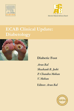
Of all lower extremity amputations, 40–70% are related to diabetes. In most studies, the incidence of lower leg amputation is estimated to be 5–25/100,000 inhabitants/year: among people with diabetes the number is 6–8/1,000. Lower extremity amputations are usually preceded by a foot ulcer in people with diabetes. The most important factors related to the development of these ulcers are peripheral neuropathy, foot deformities, minor foot trauma, and peripheral vascular disease. The spectrum of foot lesions varies in different regions of the world due to differences in socioeconomic conditions, standards of foot care and quality of footwear.
This clinical update is designed to address this condition in a comprehensive way to help the reader take important questions while managing the patient with supportive typical clinical scenarios, with which all readers will be able to identify. Thus it provides an excellent opportunity to widen one’s perspective in this area.
Table of Contents
| Section Title | Page | Action | Price |
|---|---|---|---|
| Front Cover\r | Front Cover | ||
| Front Matter\r | ia | ||
| Diabetic Peripheral\rNeuropathy and\rDiabetic Foot Ulcer\r | 5a | ||
| Copyright | ie | ||
| About the Authors | if | ||
| Contents | ig | ||
| ECAB Clinical Update InformationDiabetic Foot | i | ||
| ELSEVIER CLINICAL ADVISORY BOARD (ECAB)INDIA | i | ||
| STATEMENT OF NEED | i | ||
| DIABETIC FOOT | ii | ||
| TARGET AUDIENCE | ii | ||
| EDUCATIONAL OBJECTIVES | ii | ||
| ACCREDITATION INFORMATION | ii | ||
| DISCLAIMER | ii | ||
| DISCLOSURE OF UNLABELED USES | iii | ||
| DISCLOSURE OF FINANCIAL RELATIONSHIPSWITH ANY COMMERCIAL INTEREST | iii | ||
| RESOLUTION OF CONFLICT OF INTEREST | iii | ||
| CONTENT DEVELOPMENT COMMITTEE | iv | ||
| ENQUIRIES | iv | ||
| Introduction | 1 | ||
| Diabetic PeripheralNeuropathy andDiabetic Foot Ulcer | 5a | ||
| ABSTRACT: | 5a | ||
| KEYWORDS: | 5a | ||
| INTRODUCTION | 5 | ||
| EPIDEMIOLOGY | 7 | ||
| NATURAL HISTORY | 7 | ||
| PATHOLOGY | 8 | ||
| Myelinated Fibers | 8 | ||
| Unmyelinated Fibers | 10 | ||
| DEFINITIONS AND CLASSIFICATION | 10 | ||
| Rapidly Reversible Hyperglycemic Neuropathy | 13 | ||
| Acute Painful Neuropathy of Poor Glycemic Control | 14 | ||
| Acute Painful Neuropathy of Rapid Glycemic Control(“Insulin Neuritis”) | 14 | ||
| Mononeuropathies | 15 | ||
| Proximal Motor Neuropathies | 15 | ||
| Chronic Sensory Motor Neuropathy(Chronic Distal Symmetrical Neuropathy) | 18 | ||
| INVESTIGATION OF NEUROPATHY | 19 | ||
| Clinical Measures | 19 | ||
| Quantitative Sensory Testing | 19 | ||
| Electrophysiology | 20 | ||
| Morphologic Assessment | 20 | ||
| TREATMENT OPTIONS | 20 | ||
| Current Treatments | 21 | ||
| Treatment | 21 | ||
| Management of Distal Symmetrical Polyneuropathy | 21 | ||
| Management Aimed at Symptoms | 22 | ||
| Pain Control | 22 | ||
| Management of Disabling Painful Neuropathy NotResponding to Pharmacological Treatment | 23 | ||
| Potential Future Therapies | 24 | ||
| Aldose Reductase Inhibitors | 24 | ||
| 24 | |||
| Nerve Growth Factor | 25 | ||
| New Treatments for Autonomic Dysfunction | 25 | ||
| DIABETIC FOOT ULCER:INTRODUCTION | 25 | ||
| PATHOGENESIS | 26 | ||
| Charcot Foot | 26 | ||
| Classification | 27 | ||
| Clinical Presentation | 27 | ||
| Diagnosis: Diagnostic Procedures for DFU89 | 28 | ||
| ALTERNATIVES TO CONTRAST ANGIOGRAPHY | 29 | ||
| Prevention and Management of Foot Ulcers in Diabetes | 41 | ||
| Why do Patients of Diabetes get Foot Problems? | 44 | ||
| Evaluation Of Diabetic Neuropathy | 45 | ||
| Effects of Peripheral Neuropathy | 46 | ||
| Sudomotor Dysfunction | 46 | ||
| Arteriovenous Shunting | 47 | ||
| Development of Arteriovenous Shunts | 47 | ||
| Effects of AVS on Diabetic Foot | 47 | ||
| Contribution of Nails to the Development of Diabetic Foot Ulcers | 48 | ||
| Painless-Painful Foot Syndrome in Diabetes | 49 | ||
| Peripheral Vascular Disease in Diabetes | 49 | ||
| Pathogenetic Contribution of Vasculopathy to the Development of Neuropathy | 51 | ||
| Infection and Related Issues | 51 | ||
| Diabetes Mellitus as an Immunocompromised State | 53 | ||
| Failure of Diabetics to Control the Spread | 53 | ||
| Management of Infected Diabetic Foot | 54 | ||
| How Early Conservative Amputation Should Be Done? | 55 | ||
| Concept of Foot Spaces43 | 55 | ||
| Measures to Prevent Worsening of Polybacterial Foot Infections | 56 | ||
| Assessment | 56 | ||
| Clinical | 56 | ||
| Laboratory | 57 | ||
| Assessment of Comorbidities | 57 | ||
| Imaging Investigations (Likely Findings) | 58 | ||
| Treatment | 58 | ||
| Guidelines for the Usage of Antibiotics | 58 | ||
| Non-limb-threatening Infections | 58 | ||
| Oral | 58 | ||
| Limb-threatening Infections | 59 | ||
| Parenteral | 59 | ||
| Life-threatening Infections | 59 | ||
| Other Useful Antibiotics | 59 | ||
| Types of the Procedures | 59 | ||
| Principle | 59 | ||
| Draining Plantar Spaces | 59 | ||
| About the First Toe | 60 | ||
| First Toe Disarticulation | 61 | ||
| A Special Consideration for Trans-metatarsal Amputation | 61 | ||
| Lesser Toe Amputation | 61 | ||
| Ray Amputation | 62 | ||
| Syme's Amputation | 62 | ||
| Wound Care | 63 | ||
| Off-loading | 63 | ||
| Rehabilitation | 63 | ||
| Prevention of Recurrence | 64 | ||
| Factors Deciding the Level of Amputation | 64 | ||
| Factors Influencing the Clinical Outcome | 64 | ||
| Time of Procedure | 64 | ||
| Footwear in Diabetes | 65 | ||
| Footwear in General | 65 | ||
| Hawaii Chappals with Toe-straps | 65 | ||
| Objectives of Diabetic Footwear49 | 66 | ||
| Relief of Excessive Plantar Pressure | 66 | ||
| Reduction of Shock | 66 | ||
| Reduction of Shear (Frictional Forces) | 66 | ||
| Accommodation of Minimal Deformity | 66 | ||
| Stabilization of Deformity | 66 | ||
| Preventing Recurrence of Ulcer | 66 | ||
| Characteristics of Ideal Diabetic Footwear50 | 66 | ||
| General Guidelines for Size, Length and Fitment50 | 67 | ||
| A Classification of the Diabetic Footwear | 68 | ||
| Comfortable Pressure Reducing Walking Footwear | 68 | ||
| Prophylactic Walking Footwear | 68 | ||
| Custom-moulded Shoe | 68 | ||
| Rigid Rocker Bottoms | 68 | ||
| Roller Bottoms (Sole or Bar) | 68 | ||
| Therapeutic Off-loading Measures for Faster Healing of Ulcers | 68 | ||
| Choosing Footwear for Different Clinical States | 69 | ||
| Normal Response to Monofilament Test | 69 | ||
| With Partially Healed or Non-healing Planter Ulcer by Reducing Pressure | 70 | ||
| With Foot Deformity | 70 | ||
| Partially Amputated Foot | 70 | ||
| Rocker Soles49 | 71 | ||
| How Frequently Should Footwear be Changed?50 | 71 | ||
| How to Protect High-Pressure Areas in Foot with Footwear?51 | 71 | ||
| Assessing the Footwear Quality69 | 72 | ||
| Qualities Required at the Component and Product Level | 72 | ||
| Insole/Foot Bed | 72 | ||
| Soles | 73 | ||
| Heel Height | 74 | ||
| Pressure Socks | 74 | ||
| How Diabetic Foot Ulcer Should be Prevented and Managed in Indian Setting | 74 | ||
| Detecting Foot at Risk | 74 | ||
| Causes of Infections | 74 | ||
| History | 75 | ||
| Examination | 75 | ||
| Examination of Ulcer with the Help of a Trained/Trainable Paramedic | 78 | ||
| Surgical Management of an Ulcer even in the PHC Level-Probe and Explore | 79 | ||
| Footwear | 79 | ||
| Referral to Higher Level of Healthcare | 80 | ||
| Case Studies Prevention and Management of Foot Ulcers in Diabetes | 88 | ||
| Case Study 1 | 88 | ||
| Case Study 2 | 91 | ||
| Case Study 3 | 92 | ||
| Case Study 4 | 93 | ||
| Case Study 5 | 95 | ||
| Case Study 6 | 97 | ||
| Case Study 7 | 98 | ||
| Antibiotics in Diabetic Foot Infection | 100 | ||
| Global Update | 102 | ||
| Update in Indian Context | 103 | ||
| Personal clinical experience/management | 106 | ||
| Indianized algorithm/treatment plan | 112 | ||
| Conclusions | 113 | ||
| Case Studies Antibiotics in Diabetic Foot Infection | 120 | ||
| Case Study 1 | 120 | ||
| History | 120 | ||
| Examination | 120 | ||
| Management | 120 | ||
| Discussion | 121 | ||
| Case Study 2 | 121 | ||
| History | 121 | ||
| Examination | 121 | ||
| Management | 122 | ||
| Discussion | 122 | ||
| Case Study 3 | 122 | ||
| History | 122 | ||
| Examination | 122 | ||
| Management | 123 | ||
| Discussion | 124 | ||
| Summary | 125 | ||
| Forthcoming Books | 129 |
