Additional Information
Book Details
Abstract
The use of ultrasound in emergency medicine has proved invaluable in answering very specific, time-critical questions, such as the presence of an abdominal aortic aneurysm, or of blood in the abdomen after trauma. Unlike other imaging modalities (e.g. CT scan) it is a rapid technique that can be brought to the patient with ease.
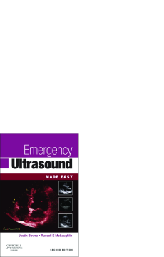
This book, Emergency Ultrasound Made Easy, is accessible and easy to use in an emergency. It is aimed mainly at specialists and trainees in emergency medicine, surgery and intensive care; but its broad scope (e.g. rapid diagnosis of DVT) makes it an invaluable addition to the library of any doctor with an interest in ultrasound, whether in primary care or the hospital setting.
- A pocket-sized and practical guide to the appropriate use of ultrasound in the emergency department.
- Designed to be used in an urgent situation (e.g. a shocked trauma patient).
- Written by team of international leading experts.
This Second Edition has been comprehensively revised and updated to reflect the major advances in the practice of bedside ultrasound, and reflects the pioneering efforts of individual clinicians and the high-quality portable machines now available. This edition still firmly adheres to the principles of only using ultrasound where it adds value and only asking simple questions that may be readily addressed using ultrasound.
Table of Contents
| Section Title | Page | Action | Price |
|---|---|---|---|
| Front cover | cover | ||
| Emergency Ultrasound Made Easy | i | ||
| Copyright page | iv | ||
| Table of Contents | v | ||
| Contributors | vii | ||
| Preface | viii | ||
| Acknowledgements | ix | ||
| Abbreviations | x | ||
| 1 Introduction | 1 | ||
| What is ultrasound? | 1 | ||
| What is emergency US? | 1 | ||
| What it isn’t (you are not a radiologist!) | 1 | ||
| First considerations | 2 | ||
| The clinical question to be answered | 2 | ||
| Limitations of emergency US | 2 | ||
| Operator and technical limitations | 3 | ||
| Will a scan change management in the emergency department (ED)? | 3 | ||
| 2 How ultrasound works | 5 | ||
| What is ultrasound? | 5 | ||
| Types of US | 5 | ||
| Producing the image | 5 | ||
| The transducer | 8 | ||
| Orientation | 9 | ||
| The keyboard | 10 | ||
| Gain | 10 | ||
| Time gain compensation | 10 | ||
| Depth | 11 | ||
| Focus/position | 11 | ||
| Freeze | 11 | ||
| Artefacts | 11 | ||
| Acoustic enhancement and acoustic windows (Fig. 2.9) | 12 | ||
| Acoustic shadowing | 12 | ||
| Edge shadows | 12 | ||
| Mirror image (Fig. 2.12) | 13 | ||
| Reverberation (Fig. 2.13) | 15 | ||
| Handy hints | 15 | ||
| 3 Abdominal aorta | 17 | ||
| The question: is there an abdominal aortic aneurysm? | 17 | ||
| Why use ultrasound? | 17 | ||
| Clinical picture | 17 | ||
| Before you scan | 19 | ||
| The technique and views | 19 | ||
| Patient’s position | 19 | ||
| Probe and scanner settings | 19 | ||
| Probe placement and landmarks | 19 | ||
| Essential views | 22 | ||
| Handy hints | 22 | ||
| What US can tell you | 23 | ||
| What US can’t tell you | 23 | ||
| Now what? | 25 | ||
| 4 Focused assessment with sonography in trauma (FAST) and extended FAST (EFAST) | 29 | ||
| The question: is there free fluid? | 29 | ||
| Why use ultrasound? | 29 | ||
| Clinical picture | 30 | ||
| Cautions and contraindications | 30 | ||
| Before you scan | 31 | ||
| Technique and views | 31 | ||
| Patient’s position | 31 | ||
| Probe and scanner settings | 31 | ||
| The five views (Fig. 4.1) | 31 | ||
| Extra views | 39 | ||
| Essential views | 39 | ||
| Handy hints | 40 | ||
| What FAST can tell you | 42 | ||
| What FAST can’t tell you | 42 | ||
| Now what? | 42 | ||
| 5 Lung and thorax | 43 | ||
| How can lung ultrasound help me? | 43 | ||
| Why use US? | 43 | ||
| Clinical picture | 44 | ||
| Cautions and contraindications | 44 | ||
| Technique and views | 44 | ||
| Patient’s position | 44 | ||
| Probe and scanner settings | 45 | ||
| The views | 45 | ||
| What to look for | 46 | ||
| Normal lung | 46 | ||
| PTX | 47 | ||
| Pleural fluid | 50 | ||
| Lung rockets | 51 | ||
| Alveolar consolidation | 53 | ||
| Handy hints and pitfalls | 54 | ||
| What lung US can help tell you | 55 | ||
| What lung US can’t tell you | 55 | ||
| Now what? | 55 | ||
| 6 Focused echocardiography and volume assessment | 57 | ||
| Why use ultrasound? | 57 | ||
| Focused versus comprehensive echo | 57 | ||
| Transthoracic versus transoesophageal echocardiography | 57 | ||
| Classic haemodynamic patterns | 58 | ||
| Diagnosis | 58 | ||
| Intervention | 58 | ||
| Scanning the patient | 59 | ||
| Preparation | 59 | ||
| Patient’s position | 59 | ||
| Probe and scanner settings | 59 | ||
| Cardiac scan | 60 | ||
| Normal anatomy | 60 | ||
| Windows | 63 | ||
| Pericardium | 65 | ||
| LV size | 66 | ||
| LV contractility | 67 | ||
| RV size and contractility | 68 | ||
| Other obvious abnormalities | 68 | ||
| IVC: diameter and collapse | 69 | ||
| Theory | 69 | ||
| Technique | 70 | ||
| Suggested US approach to the patient with undifferentiated shock | 72 | ||
| Handy hints and pitfalls | 72 | ||
| Now what? | 73 | ||
| 7 Renal tract | 75 | ||
| Introduction | 75 | ||
| Why use US? Five good reasons | 75 | ||
| Anatomy | 76 | ||
| What US can tell you | 78 | ||
| Is the bladder full? | 78 | ||
| What size are the kidneys? | 78 | ||
| Is there hydronephrosis? (see Figs 7.7 and 7.8) | 78 | ||
| False negatives for hydronephrosis | 78 | ||
| Is the hydronephrosis acute or chronic? | 78 | ||
| Is there pyelonephritis? | 78 | ||
| Can I see a stone in the kidney or ureter? | 79 | ||
| Where can I safely place the SPC? | 80 | ||
| What US can’t tell you | 80 | ||
| The technique and views | 80 | ||
| Probe placement and landmarks | 80 | ||
| Handy hints and caveats | 84 | ||
| Now what? | 84 | ||
| 8 Gall bladder and common bile duct | 87 | ||
| Introduction | 87 | ||
| Why use US? | 87 | ||
| Anatomy | 87 | ||
| What emergency US can tell you | 88 | ||
| What emergency US can’t tell you | 89 | ||
| The technique and views | 89 | ||
| Patient position, probe and scanner settings | 89 | ||
| Probe placement and landmarks | 89 | ||
| Handy hints and caveats | 95 | ||
| Now what? | 96 | ||
| 9 Early pregnancy | 97 | ||
| Introduction | 97 | ||
| Ectopic pregnancy | 97 | ||
| Why use US? | 98 | ||
| What emergency US can tell you | 100 | ||
| What emergency US can’t tell you | 100 | ||
| The role of βHCG | 100 | ||
| Clinical picture | 101 | ||
| Before you scan | 102 | ||
| The technique and views: TA scan | 102 | ||
| Patient position | 102 | ||
| Probe and scanner settings | 102 | ||
| Probe placement and landmarks | 102 | ||
| Essential views and findings | 103 | ||
| Handy hints | 108 | ||
| Now what? | 109 | ||
| 10 Ultrasound-guided procedures | 111 | ||
| Why use ultrasound? | 111 | ||
| Probe sterilization | 111 | ||
| Central venous cannulation | 114 | ||
| Anatomy | 114 | ||
| Which technique? | 115 | ||
| ‘Static’ technique | 115 | ||
| Real-time in-plane and out-of-plane techniques | 115 | ||
| CVC using real-time US | 116 | ||
| Preparation | 116 | ||
| Patient’s position | 116 | ||
| Probe and scanner settings | 116 | ||
| ‘Out-of-plane’ or transverse technique | 116 | ||
| ‘In-plane’ or longitudinal section technique: | 120 | ||
| Handy hints and pitfalls | 121 | ||
| Thoracocentesis, pericardiocentesis and paracentesis | 123 | ||
| Anatomy | 123 | ||
| Preparation | 125 | ||
| Patient’s position | 125 | ||
| Probe and scanner settings | 125 | ||
| Probe placement and landmarks | 125 | ||
| Needle placement | 125 | ||
| Thoracocentesis | 125 | ||
| Pericardiocentesis | 125 | ||
| Paracentesis | 126 | ||
| Handy hints and pitfalls | 126 | ||
| What US can tell you | 126 | ||
| What US can’t tell you | 126 | ||
| Complications of draining effusions | 126 | ||
| Suprapubic catheterization | 127 | ||
| Lumbar puncture | 128 | ||
| Technique | 128 | ||
| Handy hints and pitfalls | 131 | ||
| 11 Nerve blocks | 133 | ||
| Why use ultrasound? | 133 | ||
| Which blocks? | 133 | ||
| US appearance | 134 | ||
| Probe and scanner settings | 136 | ||
| Technique | 136 | ||
| Screening exam | 136 | ||
| Preparation | 136 | ||
| Sterile technique | 138 | ||
| TS view of nerve | 138 | ||
| In-plane needle insertion | 138 | ||
| Notes on specific nerve blocks | 142 | ||
| Interscalene block (brachial plexus) | 142 | ||
| Supraclavicular block (brachial plexus) | 143 | ||
| Axillary block | 145 | ||
| Median nerve block | 145 | ||
| Ulnar nerve block | 145 | ||
| Radial nerve block | 146 | ||
| Femoral block | 146 | ||
| Handy hints and pitfalls | 150 | ||
| 12 Deep vein thrombosis | 153 | ||
| The question: is there a deep vein thrombosis? | 153 | ||
| Why use compression ultrasound? | 153 | ||
| Anatomy (Fig. 12.1) | 154 | ||
| Clinical picture | 155 | ||
| Before you scan | 155 | ||
| The technique and views | 156 | ||
| Patient’s position | 156 | ||
| Groin to adductor canal (Fig. 12.2) | 156 | ||
| Popliteal segment (Fig. 12.3) | 156 | ||
| Probe and scanner settings | 156 | ||
| Probe placement and landmarks | 156 | ||
| Essential views | 158 | ||
| Handy hints | 159 | ||
| What three-point compression US can tell you | 163 | ||
| What three-point compression US can’t tell you | 163 | ||
| Now what? | 163 | ||
| 13 Musculoskeletal and soft tissues | 165 | ||
| The questions | 165 | ||
| Paediatric hip effusion | 165 | ||
| Why use US? | 165 | ||
| Clinical picture | 165 | ||
| Before you scan | 166 | ||
| The technique and views | 166 | ||
| Patient’s position | 166 | ||
| Probe and scanner settings | 167 | ||
| Probe placement and landmarks | 167 | ||
| Arthrocentesis | 168 | ||
| Essential views | 169 | ||
| Handy hints | 169 | ||
| What US can tell you | 169 | ||
| What US can’t tell you | 169 | ||
| Now what? | 169 | ||
| Soft tissue infections | 170 | ||
| Why use US? | 171 | ||
| The technique and views | 172 | ||
| Normal anatomy | 172 | ||
| US appearances of pathology | 172 | ||
| What US can tell you | 174 | ||
| What US can’t tell you | 174 | ||
| Shoulder dislocation | 174 | ||
| Why use US? | 175 | ||
| Anatomy | 175 | ||
| The technique and views | 175 | ||
| US appearances | 176 | ||
| Essential views | 176 | ||
| Handy hints | 176 | ||
| What US can tell you | 176 | ||
| What US can’t tell you | 177 | ||
| Fracture diagnosis | 177 | ||
| Why use US? | 177 | ||
| The technique and views (distal radius and ulna) | 177 | ||
| US appearances | 177 | ||
| Handy hints | 177 | ||
| What US can tell you | 181 | ||
| What US can’t tell you | 181 | ||
| 14 Soft-tissue foreign bodies | 183 | ||
| The question: is there a foreign body? | 183 | ||
| Why use ultrasound? | 183 | ||
| Clinical picture | 183 | ||
| The technique and views | 183 | ||
| Patient’s position | 183 | ||
| Probe and scanner settings | 183 | ||
| Probe placement and landmarks for FB localization | 185 | ||
| FB removal | 186 | ||
| Handy hints | 186 | ||
| What US can tell you | 186 | ||
| What US can’t tell you | 187 | ||
| Now what? | 187 | ||
| 15 Emergency ultrasound in combat/austere settings | 189 | ||
| Why use ultrasound in this setting? | 189 | ||
| Scope of resuscitative US in austere/combat settings | 189 | ||
| Handy hints and pitfalls | 189 | ||
| Before you scan | 190 | ||
| Airway and breathing | 190 | ||
| Airway assessment with US | 190 | ||
| Rationale | 190 | ||
| Pitfalls | 190 | ||
| Breathing assessment with US | 191 | ||
| 16 Conclusion | 193 | ||
| Audit/quality control/training | 193 | ||
| Audit and quality control | 193 | ||
| Training | 193 | ||
| Managerial | 194 | ||
| Research and future directions | 194 | ||
| Appendix 1 | 197 | ||
| Useful paperwork: logbook sheet | 197 | ||
| Sample ED ultrasound log | 197 | ||
| Appendix 2 | 199 | ||
| Useful organizations | 199 | ||
| United States of America | 199 | ||
| Australasia | 199 | ||
| United Kingdom | 199 | ||
| Appendix 3 | 201 | ||
| Further reading | 201 | ||
| Index | 203 | ||
| A | 203 | ||
| B | 203 | ||
| C | 204 | ||
| D | 204 | ||
| E | 205 | ||
| F | 205 | ||
| G | 206 | ||
| H | 207 | ||
| I | 207 | ||
| K | 207 | ||
| L | 207 | ||
| M | 208 | ||
| N | 208 | ||
| O | 208 | ||
| P | 208 | ||
| Q | 209 | ||
| R | 209 | ||
| S | 210 | ||
| T | 210 | ||
| U | 211 | ||
| V | 211 | ||
| W | 211 | ||
| Y | 211 | ||
| Z | 211 |
