Additional Information
Book Details
Abstract
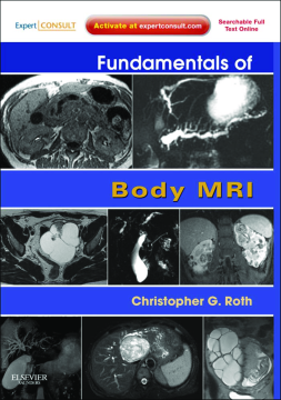
Fundamentals of Body MRI—a new title in the Fundamentals of Radiology series—explains and defines key concepts in body MRI so you can confidently make radiologic diagnoses. Dr. Christopher G. Roth presents comprehensive guidance on body imaging—from the liver to the female pelvis—and discusses how physics, techniques, hardware, and artifacts affect results. This detailed and heavily illustrated reference will help you effectively master the complexities of interpreting findings from this imaging modality.
- Master MRI techniques for the entirety of body imaging, including liver, breast, male and female pelvis, and cardiovascular MRI.
- Avoid artifacts thanks to extensive discussions of considerations such as physics and parameter tradeoffs.
- Grasp visual nuances through numerous images and correlating anatomic illustrations.
Table of Contents
| Section Title | Page | Action | Price |
|---|---|---|---|
| Front cover | cover | ||
| Inside front cover | ifc1 | ||
| Fundamentals of Body MRI | i | ||
| Copyright page | iv | ||
| Dedication | v | ||
| Contributor | vii | ||
| Preface | ix | ||
| Acknowledgments | xi | ||
| Table of Contents | xiii | ||
| ONE Introduction to Body MRI | 1 | ||
| Magnetic Resonance Imaging: What is the Objective? | 1 | ||
| Magnetism: How is the Human Body Magnetized? | 1 | ||
| The Components | 1 | ||
| The Magnet | 1 | ||
| Rf System | 3 | ||
| The Gradient System | 4 | ||
| The Receiver System | 6 | ||
| K Space and the Fourier Transform | 7 | ||
| Operator’s Console | 8 | ||
| Pulse Sequences | 9 | ||
| Tissue Contrast | 11 | ||
| The Pulse Sequence Scheme | 15 | ||
| Optimizing Body MRI | 21 | ||
| Motion | 21 | ||
| Susceptibility Artifact | 27 | ||
| MRI Safety | 28 | ||
| Summary | 33 | ||
| References | 34 | ||
| TWO MRI of the Liver | 35 | ||
| Introduction | 35 | ||
| Technique | 35 | ||
| Interpretation | 45 | ||
| Normal Features | 50 | ||
| Focal Lesions | 51 | ||
| Cystic Lesions | 53 | ||
| Developmental Lesions | 54 | ||
| Simple Hepatic Cyst | 54 | ||
| Bile Duct Hamartoma | 54 | ||
| Caroli’s Disease | 54 | ||
| Cavernous Hemangioma | 56 | ||
| Neoplastic Lesions | 61 | ||
| Biliary Cystadenoma (-adenocarcinoma) | 61 | ||
| Infectious Lesions | 61 | ||
| Echinococcal Cyst | 61 | ||
| Pyogenic Abscess | 64 | ||
| Amebic Abscess | 64 | ||
| Fungal Abscess | 66 | ||
| Traumatic Lesions | 66 | ||
| Hematoma | 66 | ||
| Biloma | 66 | ||
| Solid (and Pseudosolid) Lesions | 67 | ||
| Hypervascular Lesions | 68 | ||
| Hepatic Adenoma | 68 | ||
| Focal Nodular Hyperplasia | 70 | ||
| Focal Transient Hepatic Intensity Difference | 72 | ||
| Cirrhotic Nodules (Prehypervascular) | 72 | ||
| Hepatocellular Carcinoma | 77 | ||
| Fibrolamellar Carcinoma | 83 | ||
| Metastases | 84 | ||
| Hypovascular Lesions | 87 | ||
| Metastases | 88 | ||
| Lymphoma | 89 | ||
| Ablated Tumors | 90 | ||
| Peripheral Cholangiocarcinoma | 91 | ||
| Lipid-Based Lesions | 97 | ||
| Hepatic Angiomyolipoma | 97 | ||
| Hepatic Lipoma | 98 | ||
| Focal Steatosis (Fatty Infiltration) | 99 | ||
| Focal Fatty Sparing | 99 | ||
| Geographic or Segmental Lesions | 99 | ||
| Primarily Enhancement Lesions | 100 | ||
| Geographic THID | 101 | ||
| Other Geographic Vascular Lesions | 101 | ||
| Signal ± Enhancement Lesions | 102 | ||
| Geographic Steatosis/Iron Deposition | 102 | ||
| Confluent Fibrosis | 103 | ||
| Intrahepatic Cholestasis | 103 | ||
| Diffuse Abnormalities | 104 | ||
| Occult (General Lack of Signal and Morphologic Changes) Processes | 104 | ||
| Primarily Signal Processes | 105 | ||
| Fatty Liver Disease | 105 | ||
| Iron Depositional Disease | 106 | ||
| Primarily Morphology Diseases | 107 | ||
| Cirrhosis | 110 | ||
| Autoimmune Hepatitis | 115 | ||
| Primary Biliary Cirrhosis | 115 | ||
| Primary Sclerosing Cholangitis | 115 | ||
| Budd-Chiari Syndrome | 117 | ||
| Liver Transplantation | 120 | ||
| References | 125 | ||
| THREE MRI of the Pancreaticobiliary System | 129 | ||
| Pancreas | 129 | ||
| Anatomy and Function | 129 | ||
| Normal Appearance | 129 | ||
| Imaging Techniques | 129 | ||
| Congenital/Developmental Anomalies of the Pancreas | 130 | ||
| Annular Pancreas | 130 | ||
| Pancreas Divisum | 132 | ||
| Agenesis | 132 | ||
| Diffuse Pancreatic Disorders | 133 | ||
| Lipomatosis | 133 | ||
| Pancreatitis | 134 | ||
| Acute Pancreatitis. | 134 | ||
| Chronic Pancreatitis. | 135 | ||
| Autoimmune Pancreatitis. | 135 | ||
| Groove Pancreatitis. | 144 | ||
| Hereditary Pancreatitis. | 144 | ||
| Genetic Disorders | 145 | ||
| Cystic Fibrosis | 145 | ||
| Primary (Idiopathic) Hemochromatosis | 145 | ||
| Von Hippel–Lindau Disease | 145 | ||
| Schwachman-Diamond Syndrome | 145 | ||
| Johanson-Blizzard Syndrome | 145 | ||
| Focal Pancreatic Lesions | 148 | ||
| Solid Pancreatic Lesions | 148 | ||
| Pancreatic Adenocarcinoma. | 150 | ||
| Pancreatic Neuroendocrine (Islet Cell) Tumors. | 151 | ||
| Insulinomas. | 156 | ||
| Gastrinomas. | 156 | ||
| Glucagonomas, VIPomas, and Somatostatinomas. | 156 | ||
| Nonfunctioning Islet Cell Tumors. | 158 | ||
| Pancreatic Metastases. | 158 | ||
| Other Solid Pancreatic Lesions | 160 | ||
| Acinar Cell Carcinoma. | 160 | ||
| Lymphoma. | 160 | ||
| Cystic Pancreatic Lesions | 161 | ||
| Cysts | 161 | ||
| True Cysts. | 161 | ||
| Pseudocysts. | 161 | ||
| Von Hippel–Lindau Disease. | 161 | ||
| Neoplasms | 162 | ||
| Intraductal Papillary Mucinous Neoplasms. | 162 | ||
| Serous Cystadenoma. | 166 | ||
| Mucinous Cystic Neoplasm. | 166 | ||
| Cystic Neuroendocrine (Islet Cell) Tumor. | 166 | ||
| Solid-Cystic Papillary Epithelial Neoplasm. | 170 | ||
| Gallbladder | 170 | ||
| Anatomy | 170 | ||
| Normal Appearance | 171 | ||
| Imaging Technique | 172 | ||
| Congenital/Developmental Abnormalities of the Gallbladder | 172 | ||
| Accessory Gallbladders, Ectopia, and Agenesis | 172 | ||
| Cholelithiasis | 173 | ||
| Diffuse Processes of the Gallbladder | 173 | ||
| Cholecystitis | 173 | ||
| Acute. | 173 | ||
| Chronic. | 176 | ||
| Gangrenous. | 176 | ||
| Nonspecific Edema | 176 | ||
| Adenomyomatosis | 176 | ||
| Focal Processes of the Gallbladder | 176 | ||
| Polyp | 176 | ||
| Carcinoma | 177 | ||
| Metastases | 178 | ||
| Biliary Tree | 178 | ||
| Anatomy and Normal Appearance | 178 | ||
| Imaging Techniques | 181 | ||
| Choledochal Cyst | 182 | ||
| Choledocholithiasis | 185 | ||
| Mirizzi’s Syndrome | 185 | ||
| Biliary Obstruction | 186 | ||
| Benign Etiologies | 186 | ||
| Postoperative Biliary Strictures | 186 | ||
| Inflammatory Etiologies | 187 | ||
| Cholangitis | 187 | ||
| Primary Sclerosing Cholangitis | 187 | ||
| Infectious Cholangitis | 187 | ||
| Malignant Etiologies | 188 | ||
| Cholangiocarcinoma | 188 | ||
| Ampullary Carcinoma | 193 | ||
| References | 196 | ||
| FOUR MRI of the Kidneys and Adrenal Glands | 199 | ||
| Introduction | 199 | ||
| Technique | 199 | ||
| Interpretation | 200 | ||
| Kidneys | 203 | ||
| Normal Features | 203 | ||
| Anomalies and Pseudolesions | 204 | ||
| Focal Lesions | 207 | ||
| Cystic Lesions | 209 | ||
| Simple Renal Cyst | 209 | ||
| Polycystic Diseases | 214 | ||
| Hydronephrosis | 218 | ||
| Complex Cystic Lesions | 218 | ||
| Pyogenic Renal Abscess | 218 | ||
| Cystic Renal Cell Carcinoma | 221 | ||
| Multilocular Cystic Nephroma | 222 | ||
| Renal Hematoma | 222 | ||
| Solid Lesions | 223 | ||
| Renal Cell Carcinoma | 223 | ||
| Oncocytoma | 231 | ||
| Angiomyolipoma | 231 | ||
| Urothelial Neoplasms | 232 | ||
| Renal Lymphoma | 236 | ||
| Renal Metastases | 237 | ||
| Segmental/Diffuse Lesions | 237 | ||
| Pyelonephritis | 237 | ||
| Renal Infarct | 239 | ||
| Chronic Renal Artery Stenosis | 240 | ||
| Renal Vein Thrombosis | 240 | ||
| Adrenal Glands | 242 | ||
| Normal Features | 242 | ||
| Cystic (Nonsolid) Lesions | 244 | ||
| Adrenal Cysts | 244 | ||
| Adrenal Hemorrhage | 246 | ||
| Solid Lesions | 246 | ||
| Adrenal Adenoma | 246 | ||
| Adrenal Hyperplasia | 248 | ||
| Myelolipoma | 249 | ||
| Pheochromocytoma | 249 | ||
| Metastases | 249 | ||
| Other Adrenal Malignancies | 249 | ||
| Retroperitoneum | 249 | ||
| IVC Anomalies | 252 | ||
| Retroperitoneal Fibrosis | 253 | ||
| Inflammatory Aortic Aneurysm | 256 | ||
| Retroperitoneal Lymphoma | 257 | ||
| Retroperitoneal Metastases | 257 | ||
| References | 259 | ||
| FIVE Magnetic Resonance Imaging of the Female Pelvis | 261 | ||
| Introduction | 261 | ||
| Technique | 261 | ||
| Interpretation | 265 | ||
| Uterus | 268 | ||
| Normal Features | 268 | ||
| Endometrial Pathology | 270 | ||
| Diffuse Abnormalities | 270 | ||
| Endometritis | 270 | ||
| Hormonal Factors | 270 | ||
| Tamoxifen | 271 | ||
| Focal Abnormalities | 271 | ||
| Intrauterine Adhesions | 271 | ||
| Intrauterine Device | 271 | ||
| Cesarean Section Defect | 272 | ||
| Endometrial Polyp | 272 | ||
| Endometrial Carcinoma | 273 | ||
| Pregnancy | 282 | ||
| Myometrial Disease | 282 | ||
| Focal and Diffuse Lesions | 282 | ||
| Fibroids | 282 | ||
| Adenomyosis | 294 | ||
| Myometrial Contractions | 296 | ||
| Malignant Lesions | 297 | ||
| Global Uterine Abnormalities | 298 | ||
| Cervix and Vagina | 304 | ||
| Normal Features | 304 | ||
| Cystic Lesions | 304 | ||
| Nabothian Cyst | 304 | ||
| Other Benign Cystic Lesions | 304 | ||
| Adenoma Malignum | 310 | ||
| Solid Lesions | 310 | ||
| Cervical Carcinoma | 310 | ||
| Ovaries and Adnexa | 317 | ||
| Normal Anatomy | 317 | ||
| Cystic Lesions | 321 | ||
| Water Content | 322 | ||
| Functional Ovarian Cysts | 322 | ||
| Ovarian Inclusion Cyst | 324 | ||
| Peritoneal Inclusion Cyst | 324 | ||
| Parovarian Cysts | 324 | ||
| Lipid Content | 324 | ||
| Dermoid Cyst (Mature Cystic Teratoma) | 328 | ||
| Blood Content | 329 | ||
| Endometrioma. | 329 | ||
| Functional Hemorrhagic Cyst | 338 | ||
| Hematosalpinx | 338 | ||
| Acute Lesions | 338 | ||
| Tubovarian Abscess | 338 | ||
| Ovarian Torsion | 342 | ||
| Ectopic Pregnancy | 346 | ||
| Complex Cystic and Solid Lesions | 346 | ||
| Primary | 346 | ||
| Epithelial Neoplasms | 346 | ||
| Other Primary Ovarian Neoplasms | 351 | ||
| Secondary | 359 | ||
| Miscellaneous | 359 | ||
| Pelvic Lymphoma | 359 | ||
| Aggressive Angiomyxoma | 360 | ||
| Globally Abnormal Ovaries | 360 | ||
| Polycystic Ovary Syndrome | 360 | ||
| Ovarian Hyperstimulation Syndrome | 360 | ||
| Vascular Lesions | 360 | ||
| Pelvic Arteriovenous Malformation | 364 | ||
| Pelvic Congestion Syndrome | 365 | ||
| Ovarian Vein Thrombosis | 367 | ||
| References | 367 | ||
| Index | 369 | ||
| A | 369 | ||
| B | 369 | ||
| C | 369 | ||
| D | 370 | ||
| E | 370 | ||
| F | 371 | ||
| G | 371 | ||
| H | 371 | ||
| I | 371 | ||
| J | 372 | ||
| K | 372 | ||
| L | 372 | ||
| M | 373 | ||
| N | 373 | ||
| O | 374 | ||
| P | 374 | ||
| Q | 375 | ||
| R | 375 | ||
| S | 375 | ||
| T | 375 | ||
| U | 376 | ||
| V | 376 | ||
| W | 376 | ||
| X | 376 | ||
| Y | 376 | ||
| Z | 376 |
