BOOK
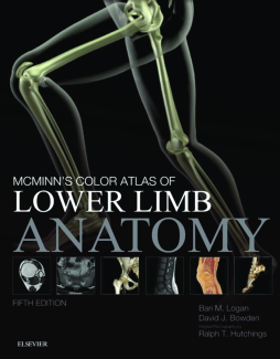
McMinn's Color Atlas of Lower Limb Anatomy E-Book
Bari M. Logan | David Bowden | Ralph T. Hutchings
(2017)
Additional Information
Book Details
Abstract
- All new and expanded ‘Imaging’ chapter to reflect what is seen in current teaching and practice
- Revised section on regional anaesthesia of the lower limb, to improve layout and reflect practice updates
Table of Contents
| Section Title | Page | Action | Price |
|---|---|---|---|
| Front Cover | cover | ||
| McMinn's Color Atlas of Lower Limb Anatomy | i | ||
| Copyright Page | iv | ||
| Table Of Contents | v | ||
| Dedication | vii | ||
| Preface | viii | ||
| Professor R. M. H. McMinn, MD (Glas), PhD (Sheff), FRCS (Eng) [b. Sept 23, 1923 – d. July 11, 2012, aged 88] | viii | ||
| McMinn’s Legacy of Illustrated Anatomy Books | ix | ||
| Acknowledgements | xii | ||
| Terminology | xiii | ||
| Preservation of Cadavers | xiii | ||
| Safety Footnote | xiii | ||
| Orientation Guides | xiv | ||
| 1 Lower limb, pelvis and hip | 1 | ||
| Lower limb survey | 2 | ||
| Bones, muscles and surface landmarks of the left lower limb, from the front | 2 | ||
| Bones, muscles and surface landmarks of the left lower limb, from behind | 4 | ||
| Bones, muscles and surface landmarks of the left lower limb, from the medial side | 6 | ||
| Bones, muscles and surface landmarks of the left lower limb, from the lateral side | 8 | ||
| Male pelvic viscera and vessels | 10 | ||
| Seen on the right side in a sagittal section, after removal of most of the peritoneum (serous membrane) | 10 | ||
| Female pelvic viscera and vessels | 12 | ||
| Seen on the right side in a sagittal section, after removal of most of the peritoneum (serous membrane) | 12 | ||
| Gluteal region | 14 | ||
| Sciatic nerve and other gluteal structures of the right side | 14 | ||
| Surface features of the right gluteal region | 15 | ||
| Left gluteal region and ischio-anal region, with gluteus maximus and gluteus medius cut through and portions reflected laterally | 16 | ||
| Right gluteal region and ischio-anal region, with most of gluteus maximus removed | 17 | ||
| Hip joint | 18 | ||
| Left hip bone and femur, with sacrum and coccyx | 18 | ||
| Axial section through the left hip joint at the level of the last, fifth segment of the sacrum, from below | 20 | ||
| 2 Thigh, knee and leg | 23 | ||
| Thigh | 24 | ||
| Front of the right thigh (female), superficial structures of the femoral triangle | 24 | ||
| Back of the right thigh (female) and gluteal region | 25 | ||
| Front of the right upper thigh (female) | 26 | ||
| Front of the right upper thigh (male) | 27 | ||
| Lower right thigh, medial side | 28 | ||
| Axial section through lower right thigh | 29 | ||
| Knee joint | 30 | ||
| Left knee joint | 30 | ||
| Left knee joint | 32 | ||
| Coronal section through the left knee joint (male), from the front | 34 | ||
| Sagittal section I through the left knee joint (female), from the left | 35 | ||
| Sagittal section II through the left knee joint (female), from the left | 36 | ||
| Sagittal section III through the left knee joint (female), from the left | 37 | ||
| Popliteal fossa and back of the knee | 38 | ||
| Popliteal fossa and back of the knee | 39 | ||
| Leg and foot survey | 40 | ||
| Muscles and superficial vessels and nerves of the left leg and foot | 40 | ||
| 3 Foot | 43 | ||
| Surface landmarks of the foot | 44 | ||
| Surface landmarks of the left foot | 44 | ||
| Surface landmarks of the left foot | 46 | ||
| Skeleton of the foot | 48 | ||
| Bones of the left foot, from above | 48 | ||
| Articulated bones of the left foot | 50 | ||
| Attachments of muscles and major ligaments to the bones of the left foot | 52 | ||
| Sesamoid and accessory bones | 53 | ||
| Articulated bones of the left foot | 54 | ||
| Bones of the left longitudinal arches, transverse tarsal joint and other joints | 56 | ||
| Foot bones | 58 | ||
| Left talus | 58 | ||
| Left talus and the lower ends of the tibia and fibula | 60 | ||
| Left talus and the lower ends of the tibia and fibula, with ligamentous attachments in the ankle region | 62 | ||
| Left talus and the lower ends of the tibia and fibula | 64 | ||
| Left talus and the lower ends of the tibia and fibula, with ligamentous attachments in the ankle region | 66 | ||
| Left calcaneus | 68 | ||
| Left navicular bone | 70 | ||
| Left cuboid bone | 70 | ||
| Articulated left cuneiform bones (medial, intermediate and lateral) | 71 | ||
| Left medial cuneiform bone | 71 | ||
| Left intermediate cuneiform bone | 71 | ||
| Left lateral cuneiform bone | 71 | ||
| Lower leg and foot | 74 | ||
| Deep fascia of the foot | 78 | ||
| Deep fascia of the right lower leg and foot, from the front and the right | 78 | ||
| Dorsum and back of the foot | 80 | ||
| Dorsum and sides of the foot | 82 | ||
| Deep nerves and vessels of the right foot, from the front and right | 84 | ||
| Deep dissection of the dorsum | 86 | ||
| Joints beneath the talus of the left foot | 86 | ||
| Sole of the foot | 88 | ||
| Plantar aponeurosis of the left foot | 88 | ||
| First layer structures | 90 | ||
| Lower leg and sole of foot | 91 | ||
| Deep medial structures and second layer from the right and slightly below | 91 | ||
| Ligaments of the foot | 96 | ||
| Ligaments of the right foot | 96 | ||
| Sections of the foot | 100 | ||
| Sagittal sections of the right foot | 100 | ||
| Sagittal sections of the right foot | 102 | ||
| Axial sections and images of the right lower leg and foot | 104 | ||
| Coronal sections of the left ankle joint and foot (in plantarflexion) | 106 | ||
| Oblique axial sections of the left foot | 109 | ||
| Coronal sections of the tarsus of the right foot | 110 | ||
| Coronal sections of the right metatarsus | 111 | ||
| Great toe | 112 | ||
| The dorsum, nail and sections of the great toe | 112 | ||
| 4 Imaging of the lower limb | 115 | ||
| Lumbar spine | 116 | ||
| Plain radiographic and CT anatomy | 116 | ||
| MRI anatomy—sagittal | 117 | ||
| MRI anatomy—axial | 118 | ||
| Pelvis | 119 | ||
| Plain radiographic anatomy | 119 | ||
| Male and female pelvis, sacrum | 120 | ||
| Developmental changes within the pelvis | 121 | ||
| MRI anatomy of the pelvis | 122 | ||
| MRI anatomy of the hip | 123 | ||
| Arterial anatomy | 124 | ||
| MRA angiographic anatomy of the pelvis and leg | 124 | ||
| Arterial anatomy of the hip | 125 | ||
| Thigh | 126 | ||
| MRI anatomy of the thigh—coronal | 126 | ||
| MRI anatomy of the thigh—axial | 127 | ||
| Knee | 128 | ||
| Plain radiographic anatomy | 128 | ||
| MRI anatomy of the knee—sagittal | 129 | ||
| MRI anatomy of the knee—coronal and axial | 130 | ||
| Arterial anatomy of the knee—DSA, CT | 131 | ||
| Arterial anatomy of the knee—CT, MRI | 132 | ||
| Lower leg | 133 | ||
| MRI anatomy of the lower leg—coronal | 133 | ||
| MRI anatomy of the lower leg—axial | 134 | ||
| Ankle | 135 | ||
| Plain radiographic anatomy | 135 | ||
| MRI anatomy of the ankle—sagittal | 136 | ||
| MRI anatomy of the ankle—axial | 137 | ||
| MRI anatomy of the ankle—coronal and ultrasound anatomy | 138 | ||
| Foot | 139 | ||
| Plain radiographic anatomy | 139 | ||
| MRI anatomy of the foot—coronal | 140 | ||
| MRI anatomy of the foot—axial and sagittal | 141 | ||
| Vascular anatomy of the foot and ankle | 142 | ||
| Paediatric anatomy | 144 | ||
| Skin | 146 | ||
| Muscles | 146 | ||
| Muscles of the gluteal region | 146 | ||
| Gluteus maximus | 146 | ||
| Gluteus medius | 146 | ||
| Gluteus minimus | 146 | ||
| Piriformis | 147 | ||
| Quadratus femoris | 147 | ||
| Obturator internus | 147 | ||
| Gemellus superior and inferior | 147 | ||
| Obturator externus | 147 | ||
| Muscles of the front of the thigh | 147 | ||
| Iliacus | 147 | ||
| Psoas major | 147 | ||
| Tensor fasciae latae | 147 | ||
| Sartorius | 147 | ||
| Rectus femoris | 147 | ||
| Vastus lateralis | 147 | ||
| Vastus medialis | 148 | ||
| Vastus intermedius | 148 | ||
| Articularis genus | 148 | ||
| Muscles of the medial side of the thigh | 148 | ||
| Pectineus | 148 | ||
| Gracilis | 148 | ||
| Adductor brevis | 148 | ||
| Adductor longus | 148 | ||
| Adductor magnus | 148 | ||
| Muscles of the back of the thigh | 148 | ||
| Biceps femoris | 148 | ||
| Semitendinosus | 148 | ||
| Semimembranosus | 148 | ||
| Muscles of the front of the leg | 149 | ||
| Tibialis anterior | 149 | ||
| Extensor hallucis longus | 149 | ||
| Extensor digitorum longus | 149 | ||
| Fibularis (peroneus) tertius | 149 | ||
| Muscle of the dorsum of the foot | 149 | ||
| Extensor digitorum brevis | 149 | ||
| Muscles of the lateral side of the leg | 149 | ||
| Fibularis (peroneus) longus | 149 | ||
| Fibularis (peroneus) brevis | 149 | ||
| Muscles of the back of the leg | 149 | ||
| Gastrocnemius | 149 | ||
| Soleus | 149 | ||
| Plantaris | 149 | ||
| Popliteus | 149 | ||
| Tibialis posterior | 149 | ||
| Flexor hallucis longus | 149 | ||
| Flexor digitorum longus | 149 | ||
| Muscles of the sole of the foot | 150 | ||
| First layer (Fig. 4) | 150 | ||
| Abductor hallucis | 150 | ||
| Flexor digitorum brevis | 150 | ||
| Abductor digiti minimi | 150 | ||
| Second layer (Fig. 5) | 150 | ||
| Quadratus plantae (flexor accessorius) | 150 | ||
| Lumbricals | 150 | ||
| Third layer (Fig. 6) | 151 | ||
| Flexor hallucis brevis | 151 | ||
| Adductor hallucis | 151 | ||
| Flexor digiti minimi brevis | 151 | ||
| Fourth layer (Fig. 7) | 151 | ||
| Dorsal interosseous (four) | 151 | ||
| Plantar interosseous (three) | 151 | ||
| Nerves | 152 | ||
| Branches of the lumbar plexus (Fig. 8) | 152 | ||
| Branches of the sacral plexus | 152 | ||
| Branches of the tibial nerve L4, L5, S1, S2, S3 (Fig. 9) | 152 | ||
| Branches of the common fibular (peroneal) nerve L4, L5, S1, S2 | 152 | ||
| Branches of the medial plantar nerve L4, L5, S1 | 153 | ||
| Branches of the lateral plantar nerve S1, S2 | 153 | ||
| Regional anaesthesia for foot and ankle | 155 | ||
| Popliteal block | 156 | ||
| Indications | 156 | ||
| Contraindications | 156 | ||
| Precautions | 156 | ||
| Anatomy | 156 | ||
| (1) Nerve stimulator-guided technique | 156 | ||
| Equipment and drugs | 156 | ||
| Procedure (Fig. 10) | 156 | ||
| Complications | 156 | ||
| (2) Ultrasound technique | 157 | ||
| Equipment and drugs | 157 | ||
| Procedure (Figs 11 and 12) | 157 | ||
| Ankle block | 158 | ||
| Indications | 158 | ||
| Contraindications | 158 | ||
| Anatomy | 158 | ||
| Equipment and drugs | 158 | ||
| Procedure | 158 | ||
| Complications | 158 | ||
| Midfoot field block | 160 | ||
| Indications | 160 | ||
| Contraindications | 160 | ||
| Anatomy | 160 | ||
| Equipment and drugs | 160 | ||
| Procedure | 160 | ||
| Complications | 160 | ||
| The common digital block | 162 | ||
| Indications | 162 | ||
| Contraindications | 162 | ||
| Equipment and drugs | 162 | ||
| Procedure (Fig. 21) | 162 | ||
| Complications | 162 | ||
| Bibliography | 162 | ||
| The lymphatic system | 165 | ||
| General key points | 165 | ||
| Lower limb lymphatics—key points | 166 | ||
| 1 Lateral caval nodes | 169 | ||
| 2 Common iliac nodes | 169 | ||
| 3 Internal iliac nodes | 169 | ||
| 4 Gluteal nodes | 169 | ||
| 5 External iliac nodes | 169 | ||
| 6 Deep inguinal nodes | 169 | ||
| 7 Popliteal nodes | 169 | ||
| 8 Superficial inguinal nodes | 169 | ||
| Arteries | 170 | ||
| Branches of the Femoral Artery | 170 | ||
| Branches of the Popliteal Artery | 170 | ||
| Branches of the Dorsalis Pedis Artery (Fig. 28) | 170 | ||
| Branches of the Medial Plantar Artery | 171 | ||
| Branches of the Lateral Plantar Artery (Fig. 29) | 171 | ||
| Index | 172 | ||
| A | 172 | ||
| B | 172 | ||
| C | 172 | ||
| D | 173 | ||
| E | 173 | ||
| F | 173 | ||
| G | 173 | ||
| H | 173 | ||
| I | 173 | ||
| J | 174 | ||
| K | 174 | ||
| L | 174 | ||
| M | 174 | ||
| N | 175 | ||
| O | 176 | ||
| P | 176 | ||
| Q | 176 | ||
| R | 176 | ||
| S | 176 | ||
| T | 177 | ||
| U | 177 | ||
| V | 177 | ||
| W | 178 | ||
| Z | 178 |
