Additional Information
Book Details
Abstract
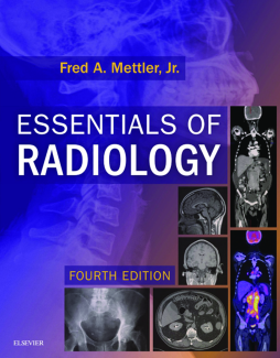
Ideal for radiology residents and medical students, as well as anyone who reads or orders radiology imaging studies, this user-friendly reference covers the basics of how to approach, read, and interpret radiological images. Using concise, step-by-step explanations and an enjoyable writing style, expert radiologist Dr. Fred A Mettler, Jr., walks you through a sequential thought process for all common indications for radiologic studies and their interpretation. Featuring thorough updates from cover to cover, this resource covers the fundamental information you need to know, as well as recent advances in the field.
- Covers which modalities to use for common suspected problems, the benefits and limitations of each modality, potential complications, clinical findings, and interpretation tips to facilitate decision-making and treatment.
- Includes normal images and common variants in primary care practice and life-threatening abnormalities for quick identification and referral – all highlighted with over 1,000 radiographic images, many in comparative panels of normal, abnormal, or correlative findings.
- Features new information throughout: more than 100 new American College of Radiology Appropriateness Criteria variants, digital breast tomosynthesis (DBT), PET/CT, new screening guidelines for colon, breast, prostate and lung cancer, new quality and safety standards, and patient and inter-professional communication.
- Incorporates today’s greater use of intermediate and advanced imaging technology, including CT, MR, and PET/CT, in addition to an emphasis on the most often-used imaging modalities such as ultrasound and plain film.
- Addresses core content of human anatomy and function/dysfunction as it relates to modern imaging.
- Features comprehensive tables of imaging indications for common problems across all body systems for quick reference.
Table of Contents
| Section Title | Page | Action | Price |
|---|---|---|---|
| Front Cover | cover | ||
| Inside Front Cover | ifc1 | ||
| Essentials of Radiology | i | ||
| Copyright Page | iv | ||
| Preface | v | ||
| Acknowledgments | vi | ||
| Table Of Contents | vii | ||
| 1 Introduction | 1 | ||
| An Approach to Image Interpretation | 1 | ||
| X-Ray | 1 | ||
| Computed Tomography | 4 | ||
| Ultrasound | 6 | ||
| Nuclear Medicine | 6 | ||
| Magnetic Resonance Imaging | 7 | ||
| Hybrid Imaging | 7 | ||
| Noninterpretative Skills, Quality, and Patient Safety | 7 | ||
| Medical Errors and Adverse Events | 9 | ||
| Suggested Textbooks and Website | 10 | ||
| General Radiology | 10 | ||
| Nuclear Medicine | 10 | ||
| Ultrasound | 10 | ||
| Computed Tomography and Magnetic Resonance | 10 | ||
| Appropriateness Criteria for Ordering Studies | 10 | ||
| 2 Head and Soft Tissues of Face and Neck | 11 | ||
| Skull and Brain | 11 | ||
| Normal Skull and Variants | 11 | ||
| Brain | 11 | ||
| Normal Anatomy | 11 | ||
| Intracranial Calcifications | 11 | ||
| Headache | 11 | ||
| Hearing Loss | 19 | ||
| Head Trauma | 20 | ||
| Suspected Intracranial Hemorrhage | 21 | ||
| Pneumocephalus | 22 | ||
| Hydrocephalus | 22 | ||
| Transient Ischemic Attack | 23 | ||
| Stroke | 23 | ||
| Intracranial Aneurysm | 24 | ||
| Primary Brain Tumors and Metastases | 25 | ||
| Vertigo and Dizziness | 26 | ||
| Suspected Intracranial Infection | 27 | ||
| Multiple Sclerosis | 27 | ||
| Dementia and Slow-Onset Mental Changes | 27 | ||
| Seizures | 27 | ||
| Psychiatric Disorders | 28 | ||
| Face | 28 | ||
| Sinuses and Sinusitis | 28 | ||
| Facial Fractures | 29 | ||
| Zygoma | 29 | ||
| Nasal | 30 | ||
| Orbital | 30 | ||
| Le Fort Fractures of the Face | 31 | ||
| Mandible | 32 | ||
| Soft Tissues of the Neck | 32 | ||
| Epiglottitis | 32 | ||
| Retropharyngeal Abscess | 32 | ||
| Subcutaneous Emphysema | 32 | ||
| Thyroid | 33 | ||
| Parathyroid | 33 | ||
| Suggested Textbooks on the Topic | 35 | ||
| 3 Chest | 36 | ||
| The Normal Chest Image | 36 | ||
| Technical Considerations | 36 | ||
| Exposure | 36 | ||
| Male Versus Female Chest | 36 | ||
| Posteroanterior Versus Anteroposterior Chest X-Rays | 36 | ||
| Upright Versus Supine Chest X-Rays | 38 | ||
| Inspiration and Expiration Chest X-Rays | 38 | ||
| Normal Anatomy and Variants | 39 | ||
| Hila and Lungs | 40 | ||
| Diaphragms | 44 | ||
| Bony Structures | 44 | ||
| Soft Tissues | 46 | ||
| Computed Tomography Anatomy | 47 | ||
| The Abnormal Chest Image | 47 | ||
| Admission, Preoperative, and Prenatal Chest X-Rays | 47 | ||
| Chest X-Ray Examinations in Occupational Medicine | 47 | ||
| Silhouette Sign | 47 | ||
| Routine Chest Radiography | 52 | ||
| Portable Chest Radiography in the Intensive Care Unit | 52 | ||
| Tubes, Wires, and Lines | 52 | ||
| Endotracheal Tube | 52 | ||
| Nasogastric Tube | 53 | ||
| Jugular or Subclavian Venous Line | 53 | ||
| Swan-Ganz or Pulmonary Artery Catheter | 54 | ||
| Pleural Tubes | 54 | ||
| Cardiac Pacers and Defibrillators | 55 | ||
| Overlying Electrocardiogram Wires and Tubes | 55 | ||
| Trauma | 57 | ||
| Acute Respiratory Illness | 57 | ||
| The Airways | 58 | ||
| Occlusion | 58 | ||
| Chronic Obstructive Pulmonary Disease | 58 | ||
| Asthma | 59 | ||
| Bronchiectasis | 60 | ||
| Atelectasis | 60 | ||
| Blebs and Bullae | 62 | ||
| Airspace Pathology | 63 | ||
| Infiltrates and Pneumonias | 63 | ||
| Community-Acquired Pneumonia in Adults | 64 | ||
| Pneumonia in Immunocompromised Patients | 66 | ||
| Aspiration Pneumonia | 67 | ||
| Tuberculosis | 67 | ||
| Fungal Lesions | 68 | ||
| Lung Abscess | 68 | ||
| Adult Respiratory Distress Syndrome | 69 | ||
| Chronic Interstitial Lung Diseases | 70 | ||
| Bioterrorist Agents | 71 | ||
| Hemoptysis | 72 | ||
| Lung Cancer and Nodules | 72 | ||
| Solitary Pulmonary Nodule | 72 | ||
| Screening for Lung Cancer | 74 | ||
| Lung Cancer | 74 | ||
| Hilar Enlargement | 75 | ||
| Metastatic Disease | 75 | ||
| Hypertension | 77 | ||
| Chest Pain and Dyspnea | 78 | ||
| Congestive Heart Failure and Pulmonary Edema | 78 | ||
| Pleural Pathology | 81 | ||
| Pneumothorax | 81 | ||
| Pneumomediastinum | 84 | ||
| Subcutaneous Emphysema | 84 | ||
| Pleural Effusions | 85 | ||
| Empyemas | 86 | ||
| Pleural Calcification and Pleural Masses | 86 | ||
| Mediastinal Lesions | 86 | ||
| Masses | 86 | ||
| Diaphragm | 92 | ||
| Diaphragmatic Rupture | 92 | ||
| Suggested Textbooks on the Topic | 92 | ||
| 4 Breast | 93 | ||
| Imaging Methods | 93 | ||
| Interpretation and Workup | 93 | ||
| Screening | 95 | ||
| Breast Pain | 96 | ||
| Suggested Textbooks on the Topic | 97 | ||
| 5 Cardiovascular System | 98 | ||
| Normal Anatomy and Imaging Techniques | 98 | ||
| Evaluation of the Cardiac Silhouette | 98 | ||
| Generalized Cardiomegaly | 98 | ||
| Left Atrial Enlargement | 102 | ||
| Left Ventricular Enlargement | 104 | ||
| Aortic Stenosis and Insufficiency | 104 | ||
| Pulmonary Artery Enlargement | 105 | ||
| Congenital Cardiac Disease | 105 | ||
| Pulmonary Embolism | 107 | ||
| Ischemic Cardiac Disease | 111 | ||
| Congestive Failure | 111 | ||
| Coronary Artery Disease and Angina | 111 | ||
| Computed Tomography Coronary Artery Screening and Calcium Scoring | 113 | ||
| Myocardial Infarction | 113 | ||
| Aorta | 114 | ||
| Anatomy and Imaging Techniques | 114 | ||
| Coarctation of the Aorta | 115 | ||
| Aortic Tears | 115 | ||
| Thoracic Aortic Aneurysms | 116 | ||
| Aortic Dissection | 116 | ||
| Peripheral Vessels | 117 | ||
| Head and Neck | 117 | ||
| Abdominal Aorta and Iliac Vessels | 118 | ||
| Renal Artery Stenosis and Hypertension | 122 | ||
| Deep Venous Thrombosis | 123 | ||
| Sudden Cold Painful Leg | 123 | ||
| Workup of Claudication | 124 | ||
| Suggested Textbooks on the Topic | 124 | ||
| 6 Gastrointestinal System | 125 | ||
| Introduction | 125 | ||
| Imaging Techniques and Anatomy | 125 | ||
| Pneumoperitoneum | 127 | ||
| Intra-Abdominal Abscesses and Fever of Unknown Origin | 129 | ||
| Feeding Tubes | 129 | ||
| Abdominal Calcifications | 130 | ||
| Acute Abdominal Pain | 133 | ||
| Abdominal Trauma | 135 | ||
| Acute Gastroenteritis | 135 | ||
| Abdominal Masses | 135 | ||
| Esophagus | 136 | ||
| Anatomy and Imaging Techniques | 136 | ||
| Dysphagia and Odynophagia | 136 | ||
| Esophageal Diverticula | 138 | ||
| Presbyesophagus | 138 | ||
| Hiatal Hernia and Gastroesophageal Reflux Disease | 138 | ||
| Foreign Bodies in the Esophagus | 140 | ||
| Strictures and Dilatation | 140 | ||
| Esophagitis and Tears | 141 | ||
| Varices | 143 | ||
| Tumors | 143 | ||
| Stomach and Duodenum | 144 | ||
| Anatomy and Imaging Techniques | 144 | ||
| Gastritis and Gastric Ulcer Disease | 144 | ||
| Duodenal Ulcers | 145 | ||
| Gastric Emptying | 146 | ||
| Gastric Dilatation or Outlet Obstruction | 146 | ||
| Liver | 147 | ||
| Cirrhosis and Alcoholic Liver Disease | 147 | ||
| Trauma | 148 | ||
| Hepatic Tumors | 148 | ||
| Hepatic Cysts | 149 | ||
| Hepatitis | 149 | ||
| Hepatic Abscess | 150 | ||
| Gallbladder and Biliary System | 150 | ||
| Anatomy and Imaging Techniques | 150 | ||
| Acute Cholecystitis | 150 | ||
| Chronic Cholecystitis and Cholelithiasis | 153 | ||
| Biliary Obstruction and Jaundice | 153 | ||
| Pancreas | 154 | ||
| Pancreatitis and Complications | 154 | ||
| Tumor | 155 | ||
| Spleen | 155 | ||
| Splenomegaly | 155 | ||
| Trauma | 155 | ||
| Small Bowel | 156 | ||
| Obstruction Versus Ileus | 156 | ||
| Benign Diseases | 158 | ||
| Tumors | 159 | ||
| Colon | 159 | ||
| Colonic Obstruction Versus Paralytic Ileus | 160 | ||
| Appendicitis | 160 | ||
| Diverticulosis and Diverticulitis | 161 | ||
| Ulcerative Colitis | 162 | ||
| Crohn Disease | 162 | ||
| Ischemic Colitis | 162 | ||
| Infectious Colitis | 163 | ||
| Pneumatosis | 163 | ||
| Polyps | 163 | ||
| Colon Carcinoma | 164 | ||
| Gastrointestinal Bleeding | 165 | ||
| Volvulus | 167 | ||
| Suggested Textbook on the Topic | 167 | ||
| 7 Genitourinary System and Retroperitoneum | 168 | ||
| Anatomy and Imaging Techniques | 168 | ||
| Kidneys | 170 | ||
| Congenital Abnormalities | 170 | ||
| Renal Cysts | 172 | ||
| Hematuria | 173 | ||
| Renal Stone Disease | 174 | ||
| Renal Failure | 176 | ||
| Pyelonephritis and Renal Infections | 177 | ||
| Renal Trauma | 179 | ||
| Renal Tumors | 179 | ||
| Renal Artery Stenosis | 180 | ||
| Renal Transplant Evaluation | 180 | ||
| Obstruction of the Renal Collecting System | 181 | ||
| The Ureter | 181 | ||
| Bladder | 184 | ||
| Anatomy and Imaging Techniques | 184 | ||
| Trauma | 184 | ||
| Incontinence | 185 | ||
| Neurologic Abnormalities | 185 | ||
| Infections | 186 | ||
| Tumors | 187 | ||
| Prostate and Scrotum | 187 | ||
| Anatomy and Imaging Techniques | 187 | ||
| Testicular or Scrotal Pain and Masses | 188 | ||
| Female Pelvis | 189 | ||
| Anatomy and Imaging Techniques | 189 | ||
| Infertility | 190 | ||
| Vaginal Bleeding | 190 | ||
| Ectopic and Intrauterine Pregnancy | 191 | ||
| Radiation During Pregnancy | 193 | ||
| Pelvic Inflammatory Disease | 194 | ||
| Pelvic Pain and Masses | 194 | ||
| Tumors | 194 | ||
| Adrenal Glands and Retroperitoneum | 195 | ||
| Adrenal Glands | 195 | ||
| Retroperitoneal Adenopathy and Neoplasms | 196 | ||
| General Textbook on the Topic | 198 | ||
| 8 Skeletal System | 199 | ||
| Introduction | 199 | ||
| Cervical Spine | 199 | ||
| Normal Anatomy | 199 | ||
| Trauma | 203 | ||
| Degenerative Changes | 209 | ||
| Chronic Neck Pain | 210 | ||
| Thoracic Spine | 210 | ||
| Anatomy | 210 | ||
| Trauma | 211 | ||
| Degenerative Changes | 213 | ||
| Lumbar Spine | 213 | ||
| Normal Anatomy and Imaging Techniques | 213 | ||
| Postsurgical Changes | 215 | ||
| Fractures | 215 | ||
| Degenerative Changes | 216 | ||
| Management of Low Back Pain | 218 | ||
| Infections and Inflammation | 218 | ||
| Spinal Neoplasms and Metastases | 219 | ||
| Unusual Lesions | 220 | ||
| Osteoporosis and Bone Mineral Measurements | 221 | ||
| Stress Fractures | 222 | ||
| Shoulder and Humerus | 222 | ||
| Normal Anatomy and Imaging | 222 | ||
| Acute Shoulder Pain | 223 | ||
| Trauma | 224 | ||
| Fractures | 224 | ||
| Acromioclavicular Separation | 224 | ||
| Dislocations | 225 | ||
| Shoulder Pain and Degenerative Changes | 225 | ||
| Tumors | 228 | ||
| Infection | 228 | ||
| Elbow | 228 | ||
| Normal Anatomy and Imaging | 228 | ||
| 9 Nonskeletal Pediatric Imaging | 291 | ||
| Suspected Child Abuse | 291 | ||
| Head | 291 | ||
| Imaging Techniques | 291 | ||
| Trauma | 291 | ||
| Headache | 291 | ||
| Childhood Seizures | 291 | ||
| Neck | 293 | ||
| Croup and Epiglottitis | 293 | ||
| Chest | 294 | ||
| Normal Anatomy and Imaging | 294 | ||
| Foreign Bodies | 294 | ||
| Tubes and Lines | 295 | ||
| Respiratory Disease in the Newborn | 298 | ||
| Bronchiolitis, Reactive Airway Disease, and Pneumonia | 299 | ||
| Asthma | 302 | ||
| Cystic Fibrosis | 302 | ||
| Pediatric Abdominal Imaging | 302 | ||
| Esophageal Fistula | 302 | ||
| Acute Gastroenteritis | 302 | ||
| Bowel Obstruction | 303 | ||
| Necrotizing Enterocolitis | 305 | ||
| Meckel Diverticulum | 306 | ||
| Neonatal Jaundice | 306 | ||
| Abdominal Masses | 306 | ||
| Appendicitis | 307 | ||
| Rectal Bleeding | 308 | ||
| Urinary Abnormalities | 308 | ||
| Fever Without Source or Unknown Origin | 309 | ||
| Suggested Textbooks and Website on the Topic | 309 | ||
| Appendix | 310 | ||
| Index | 311 | ||
| A | 311 | ||
| B | 312 | ||
| C | 312 | ||
| D | 314 | ||
| E | 314 | ||
| F | 315 | ||
| G | 316 | ||
| H | 316 | ||
| I | 317 | ||
| J | 317 | ||
| K | 317 | ||
| L | 317 | ||
| M | 318 | ||
| N | 318 | ||
| O | 319 | ||
| P | 319 | ||
| Q | 320 | ||
| R | 320 | ||
| S | 320 | ||
| T | 321 | ||
| U | 322 | ||
| V | 322 | ||
| W | 322 | ||
| X | 323 | ||
| Z | 323 | ||
| Inside Back Cover | ibc1 |
