Additional Information
Book Details
Abstract
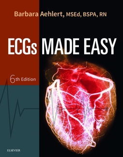
Understanding ECGs has never been easier than with ECGs Made Easy, 6th Edition! In compliance with the American Heart Association’s 2015 ECC resuscitation guidelines, Barbara Aehlert’s new edition offers clear explanations, a conversational tone, and a wealth of practice exercises to help students and professionals from a variety of medical fields learn how to accurately recognize and interpret basic dysrhythmias. Each heart rhythm covered in the book includes a sample ECG rhythm strip and a discussion of possible patient symptoms and general treatment guidelines. Other user-friendly features include: ECG Pearls with insights based on real-world experience, Drug Pearls highlighting the medications used to treat dysrhythmias, Clinical Correlation call-outs, Lead In applicatinos, Stop & Review questions, a comprehensive post-test with answers, and more. It’s everything you need to master ECG interpretation with ease!
- Clear ECG discussions highlight what you need to know about ECG mechanisms, rhythms, and heart blocks, such as: How Do I Recognize It? What Causes It? What Do I Do About It?
- Introduction to the 12-Lead ECG chapter provides all the basics for this advanced skill, including determining electrical axis, ECG changes associated with myocardial ischemia and infarction, bundle branch block, and other conditions.
- A comprehensive post-test with answers at the end of the book measures your understanding.
- ECG Pearl boxes offer useful hints for interpreting ECGs, such as the importance of the escape pacemaker.
- Drug Pearl boxes highlight various medications used to treat dysrhythmias.
- Chapter key terms focus your attention on the most important information.
- Chapter objectives tied to chapter text enable you to quickly review content that satisfies specific learning objectives.
- NEW! 38 New cardiac rhythm strips have been added to the book for a total of 260 practice strips.
- NEW! AHA compliance ensures the book reflects the American Heart Association’s 2015 ECC resuscitation guidelines.
- NEW! Lead In boxes cover ECG principles, practical applications, indications, techniques, and interpretation.
- NEW! Expanded coverage of ambulatory monitoring provides more in-depth guidance in this critical area.
Table of Contents
| Section Title | Page | Action | Price |
|---|---|---|---|
| Front Cover | Cover | ||
| ECGs MADE EASY | i | ||
| Copyright | ii | ||
| Preface to the Sixth Edition | iii | ||
| Acknowledgments | iv | ||
| Dedication | v | ||
| Reviewers for the Sixth Edition | vi | ||
| About the Author | vii | ||
| Contents | viii | ||
| 1 - Anatomy and Physiology | 1 | ||
| LOCATION, SIZE, AND SHAPE OF THE HEART | 2 | ||
| SURFACES OF THE HEART | 2 | ||
| COVERINGS OF THE HEART | 2 | ||
| STRUCTURE OF THE HEART | 5 | ||
| Layers of the Heart Wall | 5 | ||
| CARDIAC MUSCLE | 6 | ||
| Heart Chambers | 6 | ||
| ATRIA | 7 | ||
| VENTRICLES | 7 | ||
| Heart Valves | 8 | ||
| ATRIOVENTRICULAR VALVES | 8 | ||
| SEMILUNAR VALVES | 10 | ||
| HEART SOUNDS | 10 | ||
| The Heart’s Blood Supply | 11 | ||
| CORONARY ARTERIES | 11 | ||
| Right Coronary Artery | 12 | ||
| Left Coronary Artery | 13 | ||
| Coronary Artery Dominance | 13 | ||
| ACUTE CORONARY SYNDROMES | 13 | ||
| CORONARY VEINS | 16 | ||
| The Heart’s Nerve Supply | 16 | ||
| SYMPATHETIC STIMULATION | 16 | ||
| PARASYMPATHETIC STIMULATION | 17 | ||
| BARORECEPTORS AND CHEMORECEPTORS | 17 | ||
| THE HEART AS A PUMP | 19 | ||
| Cardiac Cycle | 19 | ||
| ATRIAL SYSTOLE AND DIASTOLE | 20 | ||
| VENTRICULAR SYSTOLE AND DIASTOLE | 20 | ||
| Blood Pressure | 20 | ||
| CARDIAC OUTPUT | 22 | ||
| Stroke Volume | 22 | ||
| Heart Rate | 22 | ||
| 2 - Basic Electrophysiology | 28 | ||
| CARDIAC CELLS | 30 | ||
| Types of Cardiac Cells | 30 | ||
| Properties of Cardiac Cells | 30 | ||
| CARDIAC ACTION POTENTIAL | 30 | ||
| Polarization | 31 | ||
| Depolarization | 31 | ||
| Repolarization | 32 | ||
| Phases of the Cardiac Action Potential | 32 | ||
| Refractory Periods | 34 | ||
| CONDUCTION SYSTEM | 35 | ||
| Sinoatrial Node | 35 | ||
| Atrioventricular Node and Bundle | 37 | ||
| Right and Left Bundle Branches | 38 | ||
| Purkinje Fibers | 38 | ||
| Disorders of Impulse Formation | 39 | ||
| ABNORMAL AUTOMATICITY | 39 | ||
| TRIGGERED ACTIVITY | 39 | ||
| Disorders of Impulse Conduction | 39 | ||
| CONDUCTION BLOCKS | 39 | ||
| REENTRY | 39 | ||
| Electrodes | 41 | ||
| Leads | 42 | ||
| FRONTAL PLANE LEADS | 42 | ||
| Standard Limb Leads | 42 | ||
| Augmented Limb Leads | 43 | ||
| HORIZONTAL PLANE LEADS | 44 | ||
| Chest Leads | 44 | ||
| Right Chest Leads | 45 | ||
| Posterior Chest Leads | 45 | ||
| ALTERNATIVE LEADS | 45 | ||
| Ambulatory Cardiac Monitoring | 46 | ||
| TYPES OF AMBULATORY MONITORS | 46 | ||
| Waveforms | 48 | ||
| P WAVE | 49 | ||
| QRS COMPLEX | 50 | ||
| QRS Measurement | 51 | ||
| Abnormal QRS Complexes | 51 | ||
| QRS Variations | 51 | ||
| T WAVE | 51 | ||
| Abnormal T Waves | 52 | ||
| U WAVE | 53 | ||
| Segments | 53 | ||
| PR SEGMENT | 53 | ||
| TP SEGMENT | 53 | ||
| ST SEGMENT | 53 | ||
| Abnormal ST Segments | 55 | ||
| Intervals | 55 | ||
| Abnormal PR Intervals | 55 | ||
| QT INTERVAL | 56 | ||
| R-R AND P-P INTERVALS | 56 | ||
| Artifact | 56 | ||
| Assess Regularity | 57 | ||
| VENTRICULAR REGULARITY | 57 | ||
| ATRIAL REGULARITY | 58 | ||
| Assess Rate | 58 | ||
| METHOD 1: SIX-SECOND METHOD | 58 | ||
| METHOD 2: LARGE BOXES | 58 | ||
| METHOD 3: SMALL BOXES | 58 | ||
| Identify and Examine Waveforms | 60 | ||
| Assess Intervals and Examine Segments | 60 | ||
| PR INTERVAL | 60 | ||
| QRS DURATION | 60 | ||
| QT INTERVAL | 60 | ||
| EXAMINE ST SEGMENTS | 60 | ||
| Interpret the Rhythm | 60 | ||
| 3 - Sinus Mechanisms | 76 | ||
| INTRODUCTION | 76 | ||
| How Do I Recognize It? | 77 | ||
| SINUS RHYTHM | 77 | ||
| How Do I Recognize It? | 78 | ||
| What Causes It? | 78 | ||
| What Do I Do About It? | 79 | ||
| How Do I Recognize It? | 80 | ||
| What Causes It? | 80 | ||
| What Do I Do About It? | 81 | ||
| SINUS ARRHYTHMIA | 81 | ||
| How Do I Recognize It? | 81 | ||
| What Causes It? | 81 | ||
| What Do I Do About It? | 82 | ||
| SINOATRIAL BLOCK | 82 | ||
| How Do I Recognize It? | 82 | ||
| What Causes It? | 82 | ||
| What Do I Do About It? | 83 | ||
| SINUS ARREST | 83 | ||
| How Do I Recognize It? | 83 | ||
| What Causes It? | 83 | ||
| What Do I Do About It? | 84 | ||
| 4 - Atrial Rhythms | 102 | ||
| INTRODUCTION | 103 | ||
| ATRIAL DYSRHYTHMIAS: MECHANISMS | 103 | ||
| Abnormal Automaticity | 103 | ||
| Triggered Activity | 103 | ||
| Reentry | 104 | ||
| PREMATURE ATRIAL COMPLEXES | 104 | ||
| How Do I Recognize It? | 104 | ||
| Aberrantly Conducted Premature Atrial Complexes | 106 | ||
| Nonconducted Premature Atrial Complexes | 106 | ||
| WHAT CAUSES THEM? | 106 | ||
| WANDERING ATRIAL PACEMAKER | 107 | ||
| How Do I Recognize It? | 107 | ||
| What Causes It? | 107 | ||
| What Do I Do About It? | 107 | ||
| MULTIFOCAL ATRIAL TACHYCARDIA | 108 | ||
| How Do I Recognize It? | 108 | ||
| What Causes It? | 108 | ||
| What Do I Do About It? | 108 | ||
| SUPRAVENTRICULAR TACHYCARDIA | 108 | ||
| Atrial Tachycardias | 109 | ||
| HOW DO I RECOGNIZE IT? | 110 | ||
| WHAT CAUSES IT? | 110 | ||
| WHAT DO I DO ABOUT IT? | 111 | ||
| VAGAL MANEUVERS | 112 | ||
| SYNCHRONIZED CARDIOVERSION | 112 | ||
| Indications | 113 | ||
| Atrioventricular Nodal Reentrant Tachycardia | 113 | ||
| HOW DO I RECOGNIZE IT? | 113 | ||
| WHAT CAUSES IT? | 114 | ||
| WHAT DO I DO ABOUT IT? | 114 | ||
| HOW DO I RECOGNIZE IT? | 116 | ||
| WHAT CAUSES IT? | 116 | ||
| WHAT DO I DO ABOUT IT? | 116 | ||
| ATRIAL FLUTTER | 117 | ||
| How Do I Recognize It? | 117 | ||
| What Causes It? | 118 | ||
| What Do I Do About It? | 118 | ||
| ATRIAL FIBRILLATION | 119 | ||
| How Do I Recognize It? | 119 | ||
| What Causes It? | 121 | ||
| What Do I Do About It? | 121 | ||
| 5 - Junctional Rhythms | 141 | ||
| INTRODUCTION | 141 | ||
| PREMATURE JUNCTIONAL COMPLEXES | 142 | ||
| How Do I Recognize Them? | 142 | ||
| What Causes Them? | 143 | ||
| What Do I Do About Them? | 143 | ||
| JUNCTIONAL ESCAPE BEATS OR RHYTHM | 144 | ||
| How Do I Recognize It? | 144 | ||
| What Causes It? | 145 | ||
| 6 - Ventricular Rhythms | 165 | ||
| INTRODUCTION | 166 | ||
| PREMATURE VENTRICULAR COMPLEXES | 166 | ||
| How Do I Recognize Them? | 166 | ||
| What Causes Them? | 170 | ||
| What Do I Do About Them? | 170 | ||
| VENTRICULAR ESCAPE BEATS OR RHYTHM | 170 | ||
| How Do I Recognize It? | 170 | ||
| 7 - Atrioventricular Blocks | 194 | ||
| INTRODUCTION | 194 | ||
| FIRST-DEGREE ATRIOVENTRICULAR BLOCK | 195 | ||
| How Do I Recognize It? | 195 | ||
| What Causes It? | 196 | ||
| What Do I Do About It? | 197 | ||
| SECOND-DEGREE ATRIOVENTRICULAR BLOCKS | 197 | ||
| SECOND-DEGREE ATRIOVENTRICULAR BLOCK TYPE I | 197 | ||
| How Do I Recognize It? | 197 | ||
| What Causes It? | 198 | ||
| What Do I Do About It? | 199 | ||
| SECOND-DEGREE ATRIOVENTRICULAR BLOCK TYPE II | 199 | ||
| How Do I Recognize It? | 199 | ||
| What Causes It? | 200 | ||
| What Do I Do About It? | 200 | ||
| 2:1 ATRIOVENTRICULAR BLOCK | 200 | ||
| How Do I Recognize It? | 200 | ||
| ADVANCED SECOND-DEGREE ATRIOVENTRICULAR BLOCK | 201 | ||
| THIRD-DEGREE ATRIOVENTRICULAR BLOCK | 202 | ||
| How Do I Recognize It? | 202 | ||
| What Causes It? | 203 | ||
| What Do I Do About It? | 203 | ||
| 8 - Pacemaker Rhythms | 222 | ||
| PACEMAKER SYSTEMS | 223 | ||
| Temporary Pacemakers | 224 | ||
| TRANSVENOUS PACING | 224 | ||
| EPICARDIAL PACING | 224 | ||
| TRANSCUTANEOUS PACING | 224 | ||
| Pacing Lead Systems | 225 | ||
| PACING CHAMBERS AND MODES | 226 | ||
| Single-Chamber Pacemakers | 226 | ||
| Dual-Chamber Pacemakers | 227 | ||
| Biventricular Pacemakers | 227 | ||
| Fixed-Rate Pacemakers | 227 | ||
| Demand Pacemakers | 227 | ||
| Pacemaker Codes | 228 | ||
| PACEMAKER MALFUNCTION | 228 | ||
| Failure to Pace | 228 | ||
| Failure to Capture | 229 | ||
| Failure to Sense | 230 | ||
| ANALYZING PACEMAKER FUNCTION ON THE ECG | 230 | ||
| 9 - Introduction to the 12-Lead ECG | 241 | ||
| INTRODUCTION | 241 | ||
| LAYOUT OF THE 12-LEAD ELECTROCARDIOGRAM | 242 | ||
| VECTORS | 242 | ||
| Axis | 243 | ||
| ACUTE CORONARY SYNDROMES | 244 | ||
| Anatomic Location of a Myocardial Infarction | 246 | ||
| ANTERIOR INFARCTION | 247 | ||
| R-Wave Progression | 247 | ||
| LATERAL INFARCTION | 248 | ||
| INFERIOR INFARCTION | 248 | ||
| INFEROBASAL INFARCTION | 248 | ||
| RIGHT VENTRICULAR INFARCTION | 251 | ||
| INTRAVENTRICULAR CONDUCTION DELAYS | 254 | ||
| Structures of the Intraventricular Conduction System | 254 | ||
| Bundle Branch Activation | 254 | ||
| How Do I Recognize It? | 254 | ||
| DIFFERENTIATING RIGHT BUNDLE BRANCH BLOCK FROM LEFT BUNDLE BRANCH BLOCK | 255 | ||
| Right Bundle Branch Block | 255 | ||
| Left Bundle Branch Block | 255 | ||
| An Easier Way | 256 | ||
| EXCEPTIONS | 256 | ||
| What Causes It? | 257 | ||
| What Do I Do About It? | 257 | ||
| CHAMBER ENLARGEMENT | 257 | ||
| Atrial Abnormalities | 258 | ||
| Ventricular Abnormalities | 259 | ||
| ELECTROLYTE DISTURBANCES | 260 | ||
| Sodium | 261 | ||
| HYPERNATREMIA | 261 | ||
| HYPONATREMIA | 261 | ||
| Potassium | 261 | ||
| HYPERKALEMIA | 261 | ||
| HYPOKALEMIA | 261 | ||
| Calcium | 262 | ||
| HYPERCALCEMIA | 262 | ||
| HYPOCALCEMIA | 262 | ||
| Magnesium | 263 | ||
| HYPERMAGNESEMIA | 263 | ||
| HYPOMAGNESEMIA | 263 | ||
| ANALYZING THE 12-LEAD ELECTROCARDIOGRAM | 263 | ||
| 10 - Posttest | 278 | ||
| Index | 321 | ||
| A | 321 | ||
| B | 321 | ||
| C | 322 | ||
| D | 322 | ||
| E | 322 | ||
| F | 323 | ||
| G | 323 | ||
| H | 323 | ||
| I | 323 | ||
| J | 323 | ||
| K | 323 | ||
| L | 323 | ||
| M | 323 | ||
| N | 324 | ||
| O | 324 | ||
| P | 324 | ||
| Q | 324 | ||
| R | 324 | ||
| S | 325 | ||
| T | 325 | ||
| U | 325 | ||
| V | 325 | ||
| W | 326 |
