BOOK
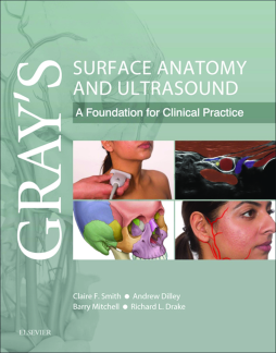
Gray’s Surface Anatomy and Ultrasound E-Book
Claire France Smith | Andrew Dilley | Barry Mitchell | Richard Drake
(2017)
Additional Information
Book Details
Abstract
A concise, superbly illustrated textbook that brings together a reliable, clear and up to date guide to surface anatomy and its underlying gross anatomy, combined with a practical application of ultrasound and other imaging modalities.
A thorough understanding of surface anatomy remains a critical part of clinical practice, but with improved imaging technology, portable ultrasound is also fast becoming integral to routine clinical examination and effective diagnosis.
This unique new text combines these two essential approaches to effectively understanding clinical anatomy and reflects latest approaches within modern medical curricula. It is tailored specifically to the needs of medical students and doctors in training and will also prove invaluable to the wide range of allied health students and professionals who need a clear understanding of visible and palpable anatomy combined with anatomy as seen on ultrasound.
Table of Contents
| Section Title | Page | Action | Price |
|---|---|---|---|
| Front Cover | cover | ||
| Inside Front Cover | ifc1 | ||
| Gray's Surface Anatomy and Ultrasound | i | ||
| Copyright Page | iv | ||
| Table Of Contents | v | ||
| Foreword | vii | ||
| Ultrasound | vii | ||
| Surface anatomy | viii | ||
| Preface | ix | ||
| About this book | x | ||
| Drawing equipment | x | ||
| Ultrasound | x | ||
| Consent | x | ||
| Expert reviewers (in order of contribution) | xi | ||
| Credits | xii | ||
| Acknowledgments | xiii | ||
| Dedications | xiii | ||
| 1 Introduction | 1 | ||
| Conceptual overview | 2 | ||
| Surface anatomy | 2 | ||
| Anatomical Position and Planes | 2 | ||
| Anatomical Terms | 3 | ||
| Movement | 3 | ||
| Fascia | 3 | ||
| Skin | 4 | ||
| Skin Color | 5 | ||
| Dermatomes and Myotomes | 6 | ||
| Natural Variation | 6 | ||
| Palpation and Percussion | 7 | ||
| Ultrasound | 7 | ||
| Ultrasound Theory | 7 | ||
| Doppler | 8 | ||
| Types of Transducer | 9 | ||
| Imaging Planes | 9 | ||
| Screen Orientation | 9 | ||
| Ergonomics | 10 | ||
| Manipulating the Transducer | 10 | ||
| Short-axis and Long-axis Views | 11 | ||
| Image terminology | 11 | ||
| Appearance of tissues | 12 | ||
| Muscle | 12 | ||
| Myofascia | 12 | ||
| Subcutaneous fat | 12 | ||
| Tendon | 12 | ||
| Hyaline cartilage | 12 | ||
| Fibrocartilage | 12 | ||
| Bone | 12 | ||
| Nerve | 12 | ||
| Blood vessels | 12 | ||
| Ligaments | 12 | ||
| Glands | 12 | ||
| Air | 12 | ||
| Fluid | 12 | ||
| 2 Thorax | 14 | ||
| Conceptual overview | 15 | ||
| Surface anatomy | 15 | ||
| Bones | 15 | ||
| Muscles | 15 | ||
| Breast | 18 | ||
| Thoracic Cavity | 18 | ||
| Pleura | 19 | ||
| Trachea | 19 | ||
| Lungs | 21 | ||
| Heart | 21 | ||
| Great vessels | 22 | ||
| Ultrasound | 26 | ||
| Anterior Muscles of the Thorax and Lungs | 26 | ||
| 3 Abdomen | 29 | ||
| Conceptual overview | 30 | ||
| Surface anatomy | 30 | ||
| Bones | 30 | ||
| Abdominal Regions | 30 | ||
| Muscles | 32 | ||
| Inguinal canal | 32 | ||
| Peritoneum | 33 | ||
| Viscera | 34 | ||
| Gastrointestinal tract | 34 | ||
| Stomach | 34 | ||
| Small intestine | 35 | ||
| Large intestine | 35 | ||
| Liver and gallbladder | 36 | ||
| Spleen | 37 | ||
| Pancreas | 37 | ||
| Kidney | 38 | ||
| Suprarenal glands | 38 | ||
| Vasculature | 38 | ||
| Ultrasound | 41 | ||
| Anterior Abdominal Musculature | 41 | ||
| Subject position | 41 | ||
| Transducer | 41 | ||
| Transducer position | 41 | ||
| Image features | 41 | ||
| Anterior abdominal wall | 41 | ||
| Anterolateral abdominal wall | 42 | ||
| Gastrointestinal Tract | 42 | ||
| Subject position | 42 | ||
| Transducer | 42 | ||
| Transducer position | 42 | ||
| Image features | 42 | ||
| Stomach | 42 | ||
| Jejunum | 42 | ||
| Ileum | 43 | ||
| Cecum and colon | 43 | ||
| Liver | 43 | ||
| Subject position | 43 | ||
| Transducer | 43 | ||
| Transducer position | 43 | ||
| Image features | 43 | ||
| Kidney | 46 | ||
| 4 Pelvis and perineum | 50 | ||
| Conceptual overview | 51 | ||
| Surface anatomy | 51 | ||
| Bones | 51 | ||
| Muscles | 51 | ||
| Viscera | 51 | ||
| Sigmoid colon | 52 | ||
| Rectum | 53 | ||
| Bladder | 54 | ||
| Ovary | 54 | ||
| Uterus | 54 | ||
| Vagina | 55 | ||
| Prostate and seminal vesicles | 55 | ||
| Perineum | 56 | ||
| Female external genitalia | 57 | ||
| Male external genitalia | 57 | ||
| Penis | 57 | ||
| Scrotum | 58 | ||
| Testis | 58 | ||
| Pregnancy | 58 | ||
| Ultrasound | 59 | ||
| Male Pelvis | 59 | ||
| Subject position | 59 | ||
| Transducer | 59 | ||
| 5 Back | 65 | ||
| Conceptual overview | 66 | ||
| Surface anatomy | 66 | ||
| Curvatures | 66 | ||
| Bones | 66 | ||
| Ligaments | 67 | ||
| Joints | 69 | ||
| Muscles | 69 | ||
| Superficial | 70 | ||
| Intermediate | 70 | ||
| Deep | 70 | ||
| The suboccipital triangle | 71 | ||
| Movements | 71 | ||
| Vertebral Canal and Spinal Nerves | 71 | ||
| Ultrasound | 77 | ||
| Subject position | 77 | ||
| Transducer | 77 | ||
| Transducer position | 77 | ||
| Image features | 77 | ||
| Cervical | 77 | ||
| Thoracic | 77 | ||
| Lumbar | 78 | ||
| 6 Upper limb | 83 | ||
| Conceptual overview | 84 | ||
| Surface anatomy | 84 | ||
| Shoulder | 84 | ||
| Bones | 84 | ||
| Muscles | 84 | ||
| Glenohumeral joint | 84 | ||
| Axilla | 87 | ||
| Arm | 89 | ||
| Bones | 89 | ||
| Muscles | 89 | ||
| Anterior compartment | 89 | ||
| Posterior compartment | 89 | ||
| Elbow joint | 89 | ||
| Cubital fossa | 90 | ||
| Forearm | 91 | ||
| Bones | 91 | ||
| Muscles | 91 | ||
| Anterior compartment | 91 | ||
| Posterior compartment | 94 | ||
| Hand | 98 | ||
| Bones | 98 | ||
| Carpal tunnel | 98 | ||
| Muscles | 100 | ||
| Movements of the thumb | 102 | ||
| Grip | 103 | ||
| Neurovascular Structures | 103 | ||
| Vasculature | 103 | ||
| Venous drainage | 104 | ||
| Nerves | 106 | ||
| Brachial plexus | 106 | ||
| Median nerve | 106 | ||
| Ulnar nerve | 106 | ||
| Musculocutaneous nerve | 108 | ||
| Axillary nerve | 108 | ||
| Radial nerve | 109 | ||
| Ultrasound | 109 | ||
| Scalene Triangle | 109 | ||
| Subject position | 109 | ||
| Transducer | 109 | ||
| Transducer position | 109 | ||
| Image features | 109 | ||
| Shoulder region | 110 | ||
| Deltoid muscle | 110 | ||
| 7 Lower limb | 126 | ||
| Conceptual overview | 127 | ||
| Surface anatomy | 127 | ||
| Gluteal region | 127 | ||
| 8 Head and neck | 165 | ||
| Conceptual overview | 166 | ||
| Surface anatomy | 166 | ||
| Head | 166 | ||
| Bones | 166 | ||
| Neurocranium | 166 | ||
| Facial skeleton | 166 | ||
| Sinuses | 168 | ||
| Mandible | 168 | ||
| Temporomandibular Joint | 168 | ||
| Muscles | 169 | ||
| Eye | 170 | ||
| External Nose | 173 | ||
| Ear | 173 | ||
| Oral Cavity | 173 | ||
| Neck | 175 | ||
| Bones | 175 | ||
| Hyoid | 175 | ||
| Vertebrae | 175 | ||
| Muscles | 176 | ||
| Triangles | 176 | ||
| Viscera | 177 | ||
| Thyroid | 177 | ||
| Larynx | 177 | ||
| Lymph | 179 | ||
| Neurovascular | 180 | ||
| Nerves | 180 | ||
| Vasculature | 180 | ||
| Ultrasound | 183 | ||
| Eye | 183 | ||
| Subject position | 183 | ||
| Transducer | 183 | ||
| Transducer position | 183 | ||
| Image features | 183 | ||
| Parotid Gland | 184 | ||
| Abbreviations | e1 | ||
| Index | 191 | ||
| A | 191 | ||
| B | 191 | ||
| C | 191 | ||
| D | 192 | ||
| E | 192 | ||
| F | 192 | ||
| G | 193 | ||
| H | 193 | ||
| I | 193 | ||
| J | 193 | ||
| K | 194 | ||
| L | 194 | ||
| M | 194 | ||
| N | 194 | ||
| O | 195 | ||
| P | 195 | ||
| Q | 195 | ||
| R | 195 | ||
| S | 196 | ||
| T | 197 | ||
| U | 197 | ||
| V | 198 | ||
| W | 198 | ||
| Z | 198 |
