Additional Information
Book Details
Abstract
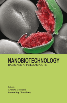
‘Nanobiotechnology, basic and applied aspects’ is expected to be of tremendous value to the group of scientists, involved in both basic and applied biology and engineering. The proposed book is a comprehensive compendium of basics of nanoscience and its application in biophysical and biomedical problems. The book describes a brief history and evolution of nanoscience in the first two chapters, which is interesting, and an enriched resource for the undergraduates of nanotechnology and biotechnology. The subsequent chapters gives an in-depth idea of different nanomaterials and their diverse biological applications such as bio-imaging, drug-development, drug-delivery, biosensors etc.. The book could also be immensely interesting for the geologists and naturalists, since it reports the occurrence of nanoparticles, which are derived from biological samples of human, and plants or of edaphic origin. In summary, the book proposed could be a reference or ready-reckoner in the undergraduate/college course-works in nanoscience and nano-biotechnology. It also gives a clear idea of different research directions in the field of nanobiotechnology.
Richard Feynman’s evolutionary idea of ‘There’s Plenty of Room at the Bottom’ is making striking innovations in our everyday life. Our body is composed of over 32 trillion of cells, which functions by virtue of nanoscale phenomena and nano-devices. The perfect orchestration of the mechanical and molecular devices at the cellular level is the most fascinating source of motivation for scientists, engaged in the research of nanoscience and biology. A picomolar volume of DNA nicely stores all the genetic information, needed to carry out cellular differentiation, programmed cell-proliferation and cell death, and the overall functioning of the living organisms. Inter and intra-cellular exchange of ions, nutrient molecules, or protein trafficking within a cell also occur via a whole system of complex guards and finely tuned molecular apertures. The biological network, which is developed in microbes, are again controlled with a molecular biosystem, distinctly different from human or higher grade of plants. These biological nanomachines inspires biologists and engineers to simulate the working-finesse of these molecular biosystems for scientific and industrials benefits and purposes.
The major goal in the nanoparticle research hence is aimed at developing new drugs for precise targeting to the disease site, effective killing of harmful microorganisms, bio-sensing in the agricultural and food industry, acute and precise bioimaging for better diagnosis of a disease or for continuous improvement of bioinstruments like microscopes etc. The list is expanding and including almost all aspects of our life. One of the major challenges in exploration into the sub-atomic size is to obtain a stable nanodevice, since at nanoscale most of the elements become highly reactive yet unstable.
Along each chapter of the book, readers will realize with amazements and wonder that at the nano-size how a materials behaves dramatically different, compared to their bulk size. It’s highly interesting to observe, how different nanomaterials (metal, non-metals, polymeric, or magnetic) have been implicated for different biological and biomedical problems. Each chapter provides an insight into the applications of nanomaterials in different biological and biomedical purposes. Undoubtedly, it is a ready reckoner for both the young and advance level researchers in the field of nanoscience and nanotechnology.
Arunava Goswami is professor in the Biological Sciences Division of Indian Statistical Institute, India. He graduated from the Tata Institute of Fundamental Research, India, and did his postdoctoral studies at Harvard Medical School, USA. Dr. Goswami was also served as a visiting faculty at Brown University, USA and Humboldt University of Berlin. He has over 50 international peer-reviewed publications, eight patents, book chapters, and more than eighty published abstracts from national and international conferences to his credit.
Samrat Roy Choudhury is currently engaged as a Research Associate in the Myeloma Institute at the University of Arkansas for Medical Sciences. He was an alumnus of Purdue University, USA and Indian Statistical Institute, India, where he pursued his postdoctoral training (2013-2016) and earned his PhD degree (Biotechnology) respectively. He has several peer-reviewed articles, patents, book chapters and a book entitled “Antibiotic resistance in E.coli and K. pneumoniae spells neonatal death (ISBN: 978-9380601328); published by LAP Lambert Academic Publishing” to his credit. Dr. Roy Choudhury has been awarded with the Best Scientist Award in Biotechnology at the 18th State Science and Technology Congress (2011) organized by the West Bengal State Govt., India.
Table of Contents
| Section Title | Page | Action | Price |
|---|---|---|---|
| Cover | Cover 1 | ||
| Front Matter | i | ||
| Half title | i | ||
| Title page | iii | ||
| Copyrights page | iv | ||
| Tables of contents | v | ||
| Preface | vii | ||
| Book Synopsis | ix | ||
| Chapter 1-6 | 1 | ||
| Chapter 1 An Introduction to The Scope of Nanoscience and Nanotechnology | 1 | ||
| What is New in Nanotechnology? | 6 | ||
| Key Elements of Nanotechnology | 8 | ||
| Possibilities of Nanotechnology | 9 | ||
| The Application of Nanotechnology to Energy Production | 9 | ||
| Nanotechnology in Agriculture and Food Science | 10 | ||
| Nanotechnology in Medicine | 12 | ||
| Nanotechnology in Electronics (Nanoelectronics) | 13 | ||
| Nanoparticles in the Atmosphere | 15 | ||
| Challenges Posed by a New Technology | 16 | ||
| Tools To Study Nanomaterials, Both Natural AndSynthetic | 16 | ||
| Rayleigh Scattering | 18 | ||
| Reflection Electron Microscope (REM) | 21 | ||
| Measuring Forces | 26 | ||
| References | 29 | ||
| Electronic references | 30 | ||
| Chapter 2 Natural Nanoparticles | 31 | ||
| Natural Nanoparticles in Living Systems | 34 | ||
| Ontogenetic Evolution | 39 | ||
| SEM Imaging | 39 | ||
| Actinomycin Treatment | 40 | ||
| Addendum | 40 | ||
| Acknowledgements | 40 | ||
| References | 41 | ||
| Chapter 3 Biological Implications of metallic Nanoparticles | 42 | ||
| 1. Synthesis | 43 | ||
| 1.1 Synthesis of Silver Nanoparticles using ElectrochemicalProcess: | 43 | ||
| 1.2 Gold Nanorods prepared by Electrochemical Method: | 43 | ||
| 1.3 Synthesis of Silver Nanoparticles with Different Shapesusing Capping Agent: | 44 | ||
| 1.4 Cylindrical Gold Nanorods Synthesis using Wet ChemicalMethod: | 44 | ||
| 1.5 Synthesis of Gold Nanorod using Seed-mediated GrowthMethod: | 44 | ||
| 1.6 Synthesis of Gold and Silver Nanoparticles usingBacteria Bacillus Subtilis: | 45 | ||
| 1.7 Using Apiin as a Reducing Agent in the Synthesis ofGold and Silver Nanoparticles: | 45 | ||
| 1.8 Biosynthesis of Silver and Gold Nanoparticles usingPhyllanthin: | 46 | ||
| 1.9 Fungus-assisted Synthesis of Silver Nanoparticles: | 46 | ||
| 1.10 Synthesis of Silver Nanoparticle with the help of PlantLeaf Extracts: | 46 | ||
| 1.11 Biosynthesis of Metal Nanoparticles using Cloves(Syzygium aromaticum) as Reducing Agent: | 47 | ||
| 1.12 Green Synthesis of Metal Nanoparticles: | 47 | ||
| 1.13 Synthesis of Water-soluble Silver Nanoparticles: | 47 | ||
| 1.14 Synthesis of Gold, Silver Nanoparticles and Gold CoreSilver Shell using Neem Leaf Broth: | 47 | ||
| 2. Application | 48 | ||
| 2.1 Biological Tagging of Antibody-conjugated GoldNanoparticles: | 48 | ||
| 2.2 Use of Gold Nanoparticles in PIC Imaging: | 49 | ||
| 2.3 Application of Silver Nanoparticles in NeuroblastomaCell as Biological Labels: | 50 | ||
| 2.4 Gold-silica Core-shell Nanorods: A Promising Materialfor Molecular Photoacoustic Imaging: | 51 | ||
| 2.5 Fluorescent Gold Nanoclusters in Biological Labeling: | 51 | ||
| 2.6 Bioconjugated Polyelectrolyte-coated GNRs as SensitiveOptical Probes for Biological Labeling: | 52 | ||
| 2.7 GNPs as Fluorescent Probes: | 52 | ||
| 2.8 Use of GNPs to Activate Enzymes and BiocatalyticProcesses: | 53 | ||
| 2.9 Multifunctional Gold Nanoparticles: | 53 | ||
| 2.10 Use of Gold Nanorods Applied in Molecularly-targetedPhotodiagnostics and Therapy: | 54 | ||
| 2.11 Development of Gold Nanoparticles-assistedColorimetric Assay for Detection of Cancer Cell: | 55 | ||
| 2.12 Using Gold Nanoparticles in Drug-delivery of PlatinumbasedAnticancer Drugs: | 55 | ||
| 2.13 Biosensor based on Gold and Silver Nanoparticles: | 56 | ||
| 2.14 Using Gold Nanoparticles for Glycation Sensing: | 56 | ||
| Reference | 57 | ||
| Chapter 4 Non-Metallic Nanoparticles &Their Biological Implications | 61 | ||
| 1. An Overview of Carbon Nanotubes (CNTs) | 62 | ||
| 1.1 Electric Arc Discharge | 62 | ||
| 1.2 Laser Ablation Method | 63 | ||
| 1.3 Chemical Vapor Deposition (CVD) | 63 | ||
| 1.4 High-pressure Carbon monoxide DisproportionationMethod (HiPco) | 64 | ||
| 1.5 Biological Implications of CNTs | 64 | ||
| 2. An Overview of Selenium Nanoparticles | 65 | ||
| 2.1 Selenium Nanoparticles on Cellulose Nanocrystals | 65 | ||
| 2.2 Selenium Nanoparticles from Sodium selonosulfate | 66 | ||
| 2.3 Selenium Nanoparticles wrapped within ChitosanPolymer | 66 | ||
| 2.4 Biosynthesis of Selenium Nanoparticles | 66 | ||
| 2.5 Biological Implications of Selenium Nanoparticles | 67 | ||
| 2.5.1 Inhibition of oxidative stress by selenium nanoparticles | 67 | ||
| 2.5.2 Selenium nanoparticles-mediated induction for mitochondrialapoptosis | 67 | ||
| 3. An Overview of Sulfur Nanoparticles | 68 | ||
| 3.1 Liquid Synthesis Method | 69 | ||
| 3.2 Cysteine Modification Method | 70 | ||
| 3.3 Wate- in-Oil Microemulsion Technique | 70 | ||
| 3.4 Biological Implications of Sulfur Nanoparticles | 70 | ||
| Reference | 71 | ||
| Chapter 5 Magnetic Nanoparticles | 75 | ||
| 1. Problems arising from Nanotherapeutics | 77 | ||
| 2. Coating of Magnetic Particle | 77 | ||
| 2.1. Chitosan | 78 | ||
| 2.2. Polyethyleneimine (PEI) | 78 | ||
| 2.3. Polyethylene glycol (PEG) | 79 | ||
| 2.4 Dextran | 79 | ||
| 2.5 Liposomes and Micelles | 80 | ||
| 3. Targeting of Magnetic Nanoparticles for Diagnosisand Detection | 80 | ||
| 3.1 Physical targeting | 80 | ||
| 3.2. Passive Targeting | 81 | ||
| 3.2.1. Enhanced permeability and retention (EPR) | 81 | ||
| 3.2.2. Size-dependant distribution of MNPs in tissues | 81 | ||
| 3.2.3. Charge-induced | 82 | ||
| 3.2.4. Reticuloendothelial system (RES) | 83 | ||
| 3.3. Active Targeting | 83 | ||
| 3.3.1. Antibodies | 83 | ||
| 3.3.2. Aptamer | 85 | ||
| 3.3.3 Peptides | 86 | ||
| 3.3.3.1. Tumor-homing peptides | 86 | ||
| 3.3.3.2. Chlorotoxin for targeting tumors of brain/neuroectodermal origin | 86 | ||
| 3.3.3.3. Bombesin-targeted CLIO contrast agents for imaging pancreaticductal adenocarcinoma | 87 | ||
| 3.3.3.5. EPPT for targeting uMUC-1 over-expressing cancer cell-lines | 87 | ||
| 3.3.3.6. LHRH-conjugated ULTRA MNP for in-vivo cancer diagnosis/imaging | 87 | ||
| 3.4. Small Molecules as Targeting Agents | 88 | ||
| 4. Conjugation Agents for MNPs | 89 | ||
| 5. Applications of MNPs | 90 | ||
| 5.1 Drug delivery | 90 | ||
| 5.2 Cancer imaging | 91 | ||
| 5.3 Molecular imaging | 91 | ||
| 5.4 Cardiovascular disease imaging | 91 | ||
| Reference | 91 | ||
| Chapter 6 Biological implications of Polymer nanocomposites | 104 | ||
| Routes to Polymer-Nanocomposites Synthesis | 106 | ||
| Characterization of Polymer Nanocomposites | 112 | ||
| Biological Implications of Polymer Nanocomposites | 113 | ||
| Conclusions | 115 | ||
| References | 115 | ||
| End Matter | 119 | ||
| Index | 119 |
