BOOK
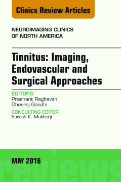
Tinnitus: Imaging, Endovascular and Surgical Approaches, An issue of Neuroimaging Clinics of North America, E-Book
Prashant Raghavan | Dheeraj Gandhi
(2016)
Additional Information
Book Details
Abstract
This issue of Neuroimaging Clinics of North America focuses on Imaging of Tinnitus. Articles will include: Neuroscience of Tinnitus; Clinical Evaluation of the Patient with Tinnitus; Arterial Abnormalities Leading to Tinnitus; Paragangliomas and Other Vascular Skull Base Tumors; Dural AV Fistulae: Imaging and Management; Venous Abnormalities Leading to Tinnitus: Imaging Evaluation; Endovascular Intervention in Venous Tinnitus: New Horizons and Future Directions; Emerging Role of Surgical Treatments in the Treatment of Tinnitus; Role of Advanced Neuroimaging and Future Directions; Imaging Interpretation of Temporal Bone Studies in a Patient with Tinnitus: A Systematic Approach; and much more!
Table of Contents
| Section Title | Page | Action | Price |
|---|---|---|---|
| Front Cover | Cover | ||
| Tinnitus: Imaging,Endovascular andSurgical Approaches | i | ||
| Copyright\r | ii | ||
| CME Accreditation Page | iii | ||
| PROGRAM OBJECTIVE | iii | ||
| TARGET AUDIENCE | iii | ||
| LEARNING OBJECTIVES | iii | ||
| ACCREDITATION | iii | ||
| DISCLOSURE OF CONFLICTS OF INTEREST | iii | ||
| UNAPPROVED/OFF-LABEL USE DISCLOSURE | iii | ||
| TO ENROLL | iii | ||
| METHOD OF PARTICIPATION | iii | ||
| CME INQUIRIES/SPECIAL NEEDS | iii | ||
| NEUROIMAGING CLINICS OF NORTH AMERICA\r\r | iv | ||
| FORTHCOMING ISSUES | iv | ||
| August 2016 | iv | ||
| November 2016 | iv | ||
| February 2017 | iv | ||
| RECENT ISSUES | iv | ||
| February 2016 | iv | ||
| November 2015 | iv | ||
| August 2015 | iv | ||
| Contributors | v | ||
| CONSULTING EDITOR | v | ||
| EDITORS | v | ||
| AUTHORS | v | ||
| Contents | vii | ||
| Foreword: Tinnitus\r | vii | ||
| Preface: Tinnitus—More Than Ringing in the Ears\r | vii | ||
| Neuroscience of Tinnitus\r | vii | ||
| Clinical Evaluation of Tinnitus\r | vii | ||
| Imaging Interpretation of Temporal Bone Studies in a Patient with Tinnitus: A Systematic Approach\r | vii | ||
| Arterial Abnormalities Leading to Tinnitus\r | vii | ||
| Venous Abnormalities Leading to Tinnitus: Imaging Evaluation\r | viii | ||
| Dural Arteriovenous Fistulae: Imaging and Management\r | viii | ||
| Paragangliomas of the Head and Neck\r | viii | ||
| Surgical Treatment of Tinnitus\x0B | viii | ||
| Endovascular Interventions for Idiopathic Intracranial Hypertension and Venous Tinnitus: New Horizons\r | ix | ||
| Advanced Neuroimaging of Tinnitus\r | ix | ||
| Foreword:\rTinnitus | xi | ||
| Preface:\rTinnitus—More Than Ringing in the Ears | xiii | ||
| Neuroscience of Tinnitus | 187 | ||
| Key points | 187 | ||
| ANATOMY OF THE AUDITORY PATHWAY | 187 | ||
| BIOCHEMICAL AND MOLECULAR TRANSDUCTION | 188 | ||
| CENTRAL NETWORK ORGANIZATION | 188 | ||
| PERCEPTION AND NONAUDITORY DIMENSIONS OF SOUND | 189 | ||
| PERIPHERAL AUDITORY SYSTEM RESPONSE TO INSULT | 189 | ||
| CENTRAL DISORDERS OF TINNITUS | 190 | ||
| CLINICAL THERAPY, EXPERIMENTAL APPROACHES, AND FINDINGS | 191 | ||
| SUMMARY | 193 | ||
| REFERENCES | 193 | ||
| Clinical Evaluation of Tinnitus | 197 | ||
| Key points | 197 | ||
| INTRODUCTION | 197 | ||
| CLINICAL EVALUATION OF TINNITUS | 198 | ||
| Basic Otologic Evaluation | 198 | ||
| History | 198 | ||
| Physical examination | 199 | ||
| Audiogram | 200 | ||
| Evaluation of subjective tinnitus | 200 | ||
| Evaluation of objective tinnitus | 202 | ||
| Patulous eustachian tube | 203 | ||
| Palatal myoclonus | 203 | ||
| Tensor tympani/stapedial myoclonus | 203 | ||
| Tinnitus from an abnormal arterial or venous somatosound | 203 | ||
| SUMMARY | 204 | ||
| REFERENCES | 204 | ||
| Imaging Interpretation of Temporal Bone Studies in a Patient with Tinnitus | 207 | ||
| Key points | 207 | ||
| BACKGROUND (EPIDEMIOLOGY, PATHOPHYSIOLOGY, AND ANATOMY: CLINICAL PERSPECTIVE) | 207 | ||
| IMAGING APPROACH | 208 | ||
| Imaging Perspective | 208 | ||
| Imaging Guidelines | 208 | ||
| DIFFERENTIAL DIAGNOSIS | 209 | ||
| VASCULAR CAUSES | 210 | ||
| Vascular Malformations | 210 | ||
| Arteriovenous fistulae | 210 | ||
| Arteriovenous malformations | 210 | ||
| Acquired Arterial Diseases | 210 | ||
| Aneurysms | 210 | ||
| Arterial dissection | 211 | ||
| Atherosclerotic stenosis | 212 | ||
| Fibromuscular dysplasia | 212 | ||
| Congenital Arterial Variants | 213 | ||
| Aberrant internal carotid artery | 213 | ||
| Persistent stapedial artery | 215 | ||
| Venous Causes | 215 | ||
| Sigmoid sinus diverticula and dehiscence | 215 | ||
| High-riding jugular bulb | 216 | ||
| INTRACRANIAL PRESSURE ABNORMALITIES | 216 | ||
| Idiopathic Intracranial Hypertension | 216 | ||
| Intracranial Hypotension | 216 | ||
| NEOPLASMS | 217 | ||
| Posterior Fossa Masses | 217 | ||
| Vestibular schwannomas | 217 | ||
| Endolymphatic sac tumors | 218 | ||
| Jugular Fossa and Middle Ear Masses | 218 | ||
| Paragangliomas | 218 | ||
| Facial nerve schwannomas | 220 | ||
| Facial nerve hemangiomas | 220 | ||
| OSSEOUS CAUSES | 221 | ||
| Otosclerosis | 221 | ||
| Paget Disease | 222 | ||
| MISCELLANEOUS CAUSES | 222 | ||
| Meniere Disease | 222 | ||
| SUMMARY | 223 | ||
| REFERENCES | 223 | ||
| Arterial Abnormalities Leading to Tinnitus | 227 | ||
| Key points | 227 | ||
| INTRODUCTION | 227 | ||
| PATHOPHYSIOLOGY OF VASCULAR PULSATILE TINNITUS | 228 | ||
| ETIOLOGIES OF PULSATILE TINNITUS | 228 | ||
| ARTERIAL CAUSES OF PULSATILE TINNITUS | 228 | ||
| Arteriosclerotic Disease | 228 | ||
| Fibromuscular Dysplasia | 228 | ||
| Arterial Dissection | 229 | ||
| Aberrant Internal Carotid Artery | 229 | ||
| Isolated Stapedial Artery | 229 | ||
| Dehiscence of the Internal Carotid Artery | 230 | ||
| Arterial Compression of the Vestibulochoclear Nerve | 231 | ||
| Cerebral Aneurysms | 232 | ||
| Increased Cardiac Output | 232 | ||
| CAUSES OF PULSATILE TINNITUS ARISING AT THE ARTERIOVENOUS JUNCTION | 232 | ||
| Paragangliomas | 232 | ||
| Other Vascular Skull Base Neoplasms | 232 | ||
| Vascular Malformations | 233 | ||
| Osseous Dysplasias | 233 | ||
| Capillary Hyperemia Involving the Temporal Bone | 233 | ||
| VENOUS CAUSES OF PULSATILE TINNITUS | 233 | ||
| DIAGNOSTIC WORK-UP OF TINNITUS | 233 | ||
| IMAGING STRATEGIES IN THE EVALUATION OF TINNITUS | 234 | ||
| NONINVASIVE IMAGING MODALITIES USED IN THE EVALUATION OF TINNITUS | 234 | ||
| INVASIVE IMAGING EVALUATION OF TINNITUS | 234 | ||
| Catheter Angiography | 234 | ||
| SUMMARY | 235 | ||
| REFERENCES | 235 | ||
| Venous Abnormalities Leading to Tinnitus | 237 | ||
| Key points | 237 | ||
| INTRODUCTION | 237 | ||
| IDIOPATHIC INTRACRANIAL HYPERTENSION | 237 | ||
| SIGMOID SINUS WALL ANOMALIES | 238 | ||
| HIGH-RIDING/DEHISCENT JUGULAR BULB | 242 | ||
| POSTERIOR FOSSA EMISSARY VEINS | 243 | ||
| SUMMARY | 244 | ||
| REFERENCES | 244 | ||
| Dural Arteriovenous Fistulae | 247 | ||
| Key points | 247 | ||
| INTRODUCTION | 247 | ||
| CLASSIFICATION | 249 | ||
| IMAGING FINDINGS | 250 | ||
| GENERAL MANAGEMENT | 250 | ||
| ENDOVASCULAR TREATMENT | 252 | ||
| EMBOLIZATION ACCESS ROUTES | 252 | ||
| Transvenous Embolization | 252 | ||
| Transarterial Embolization | 253 | ||
| EMBOLIC AGENTS | 253 | ||
| REFERENCES | 256 | ||
| Paragangliomas of the Head and Neck | 259 | ||
| Key points | 259 | ||
| INTRODUCTION | 259 | ||
| NORMAL ANATOMY AND IMAGING TECHNIQUE | 260 | ||
| Normal Anatomy | 260 | ||
| Imaging Technique | 263 | ||
| Imaging Protocols | 264 | ||
| PATHOLOGY/IMAGING FINDINGS | 267 | ||
| Pathology | 267 | ||
| Imaging Findings | 267 | ||
| Jugulartympanic paragangliomas | 267 | ||
| Carotid body paraganglioma | 272 | ||
| Preoperative Imaging | 272 | ||
| Angiography | 272 | ||
| Embolization | 272 | ||
| DIAGNOSTIC CRITERIA | 274 | ||
| DIFFERENTIAL DIAGNOSIS | 274 | ||
| PEARLS, PITFALLS, VARIANTS | 275 | ||
| WHAT THE REFERRING PHYSICIAN NEEDS TO KNOW | 275 | ||
| SUMMARY | 275 | ||
| REFERENCES | 276 | ||
| Surgical Treatment of Tinnitus | 279 | ||
| Key points | 279 | ||
| INTRODUCTION | 279 | ||
| CLINICAL EVALUATION | 280 | ||
| NONSPECIFIC TREATMENTS (IE, TREATMENTS FOR OTHER INDICATIONS WITH SECONDARY TINNITUS BENEFIT) | 281 | ||
| SURGERY FOR PRIMARY INDICATION OF TINNITUS | 282 | ||
| Subjective Tinnitus | 282 | ||
| Objective Tinnitus | 282 | ||
| SIGMOID SINUS WALL ANOMALIES | 283 | ||
| SUMMARY | 287 | ||
| REFERENCES | 287 | ||
| Endovascular Interventions for Idiopathic Intracranial Hypertension and Venous Tinnitus | 289 | ||
| Key points | 289 | ||
| BACKGROUND | 289 | ||
| VENOUS TINNITUS | 290 | ||
| IDIOPATHIC INTRACRANIAL HYPERTENSION | 290 | ||
| Mechanism of Venous Stenosis and Venous Hypertension | 290 | ||
| Clinical Management | 290 | ||
| Endovascular Treatment Considerations | 291 | ||
| OTHER CAUSES OF VENOUS TINNITUS AMENABLE TO ENDOVASCULAR INTERVENTIONS | 293 | ||
| IMAGING OF VENOUS TINNITUS | 293 | ||
| ABERRANT VENOUS ANATOMY | 293 | ||
| Advanced Neuroimaging of Tinnitus | 301 | ||
| Key points | 301 | ||
| INTRODUCTION | 301 | ||
| NEURAL CORRELATES OF TINNITUS | 301 | ||
| FUNCTIONAL MR IMAGING IN TINNITUS | 302 | ||
| ADVANCED STRUCTURAL IMAGING | 304 | ||
| MENIERE DISEASE | 304 | ||
| ADVANCED IMAGING OF DURAL ARTERIOVENOUS FISTULAE | 305 | ||
| SUMMARY | 310 | ||
| ACKNOWLEDGMENTS | 310 | ||
| REFERENCES | 310 | ||
| Index | 313 |
