Additional Information
Book Details
Abstract
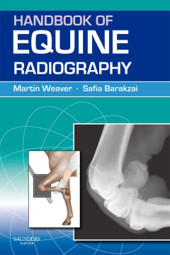
The Handbook of Equine Radiography is a practical and accessible “how-to guide to obtaining high-quality radiographs of the horse. It covers all aspects of taking radiographs of the commonly examined regions (lower limbs and skull) as well as less frequently examined areas (upper limbs, trunk).
The main part of the book consists of diagrams to illustrate the positioning of the horse and the radiography equipment. For each view a benchmark example of a normal radiograph is illustrated. The accompanying text for each radiographic view succinctly presents the most relevant aspects.
Practically orientated, and including chapters covering such key areas as radiation safety in equine radiography and patient preparation, plus a trouble-shooting section, the Handbook of Equine Radiography is an indispensable guide to practitioners in all countries engaged in equine work.
- Clear diagrams illustrate the positioning of the horse and the radiography equipment
- Contains all the information required to radiograph a horse
- Accessible to veterinary surgeons who obtain most of their radiographs in the field
Table of Contents
| Section Title | Page | Action | Price |
|---|---|---|---|
| Front cover | Cover | ||
| Title page | iii | ||
| Copyright page | iv | ||
| Table of contents | v | ||
| PART 1: PRINCIPLES OF RADIOGRAPHY | 1 | ||
| CHAPTER 1: IMAGE FORMATION | 3 | ||
| CHAPTER 2: RADIOGRAPHIC EQUIPMENT | 5 | ||
| X-ray machine | 5 | ||
| Cassettes and screens | 5 | ||
| Grids | 6 | ||
| Film labels | 6 | ||
| Cassette holders | 6 | ||
| Foot-positioning aids | 7 | ||
| Computerised radiography and direct digital radiography | 7 | ||
| CHAPTER 3: RADIATION SAFETY AND PATIENT PREPARATION | 9 | ||
| Principles of radiation safety | 9 | ||
| Radiation sources | 9 | ||
| Restricting exposure to radiation | 10 | ||
| Patient preparation | 12 | ||
| CHAPTER 4: RADIOLOGICAL INTERPRETATION AND DIAGNOSIS | 13 | ||
| PART 2: RADIOGRAPHIC PROCEDURES | 17 | ||
| CHAPTER 5: RADIOGRAPHY OF THE FOOT | 19 | ||
| Introduction | 19 | ||
| Preparation | 19 | ||
| Views | 20 | ||
| CHAPTER 6: RADIOGRAPHY OF THE PASTERN | 35 | ||
| Introduction | 35 | ||
| Preparation | 35 | ||
| Standard views | 35 | ||
| CHAPTER 7: RADIOGRAPHY OF THE FETLOCK | 41 | ||
| Introduction | 41 | ||
| Preparation | 41 | ||
| Standard views | 42 | ||
| Additional views | 48 | ||
| CHAPTER 8: RADIOGRAPHY OF THE METACARPUS AND METATARSUS | 53 | ||
| Introduction | 53 | ||
| Patient preparation | 53 | ||
| Views | 54 | ||
| CHAPTER 9: RADIOGRAPHY OF THE CARPUS | 65 | ||
| Introduction | 65 | ||
| Preparation | 65 | ||
| Views | 66 | ||
| CHAPTER 10: RADIOGRAPHY OF THE TARSUS | 79 | ||
| Introduction | 79 | ||
| Patient preparation | 79 | ||
| Views | 82 | ||
| CHAPTER 11: RADIOGRAPHY OF THE ELBOW | 95 | ||
| Introduction | 95 | ||
| Patient preparation | 95 | ||
| Views | 96 | ||
| CHAPTER 12: RADIOGRAPHY OF THE SHOULDER | 103 | ||
| Introduction | 103 | ||
| Patient preparation | 103 | ||
| Views | 104 | ||
| CHAPTER 13: RADIOGRAPHY OF THE STIFLE | 109 | ||
| Introduction | 109 | ||
| Patient preparation | 109 | ||
| Views | 110 | ||
| CHAPTER 14: RADIOGRAPHY OF THE HIP AND PELVIS | 121 | ||
| Introduction | 121 | ||
| Preparation | 121 | ||
| Views | 122 | ||
| CHAPTER 15: RADIOGRAPHY OF THE CERVICAL VERTEBRAE | 127 | ||
| Introduction | 127 | ||
| Preparation | 127 | ||
| CHAPTER 16: RADIOGRAPHY OF THE THORACOLUMBAR AND SACRAL VERTEBRAE | 133 | ||
| Introduction | 133 | ||
| Radiographic positioning | 133 | ||
| CHAPTER 17: RADIOGRAPHY OF THE SKULL | 137 | ||
| Introduction | 137 | ||
| Patient preparation | 137 | ||
| Views | 140 | ||
| CHAPTER 18: RADIOGRAPHY OF THE THORAX | 161 | ||
| Introduction | 161 | ||
| Patient preparation | 161 | ||
| Views | 162 | ||
| CHAPTER 19: RADIOGRAPHY OF THE ABDOMEN | 169 | ||
| Introduction | 169 | ||
| CHAPTER 20: ADDITIONAL RADIOGRAPHIC PROCEDURES (CONTRAST RADIOGRAPHY) | 171 | ||
| Contrast radiography of the foot | 171 | ||
| Appendix I: SUGGESTED EXPOSURE CHART | 173 | ||
| Appendix II: EXPOSURE CHART FOR YOUR PRACTICE | 177 | ||
| INDEX | 181 |
