Additional Information
Book Details
Abstract
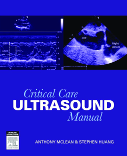
Critical Care Ultrasound Manual is a concise step-by-step guide on the assessment of ultrasounds. It trains critical care physicians in applying Rapid Assessment by Cardiac Echo (RACE) and Focused Assessment with Sonography in Trauma (FAST) to sonography principles. Animated video clips of procedures assist the reader in comprehending the content covered in the manual.
- Focus on helping readers obtain rapid practical information to assist management decisions.
- User-friendly layout.
- Explanatory diagrams, ultrasound images enhance the learning experience.
- DVD showing video clips of procedures cross-referenced in the book.
- Practical tips and cautions are highlighted in Boxes.
- MCQs on each chapter allow readers to analyse what they’ve learnt.
- The Appendices provide a checklist to assist interpretation of transthoracic echocardiogram in a systematic way, and a chapter on Doppler principles for those who wish to prepare the way for Doppler measurements.
Table of Contents
| Section Title | Page | Action | Price |
|---|---|---|---|
| Front cover | cover | ||
| Critical care ultrasound manual | i | ||
| Copyright page | iv | ||
| Table of Contents | v | ||
| Foreword | vii | ||
| Authors and Contributors | ix | ||
| Reviewers | xi | ||
| Preface | xiii | ||
| Acknowledgements | xv | ||
| 1 Basic 2D ultrasound physics | 1 | ||
| Introduction | 1 | ||
| Properties of sound waves | 1 | ||
| The ultrasound transducer | 3 | ||
| Beam formation | 3 | ||
| Reflection versus transmission | 4 | ||
| Forming an image | 5 | ||
| Types of transducers | 6 | ||
| MCQs | 8 | ||
| Chapter 1 MCQS | 8 | ||
| 2 Knobology | 9 | ||
| Introduction | 9 | ||
| Sensitivity controls | 9 | ||
| Gain | 9 | ||
| Reject | 9 | ||
| Time gain compensation | 9 | ||
| Dynamic range | 11 | ||
| Compression | 12 | ||
| Resolution controls | 13 | ||
| Lateral resolution – focus | 13 | ||
| Axial resolution – frequency | 13 | ||
| Temporal resolution – frame rate | 16 | ||
| MCQs | 17 | ||
| Chapter 2 MCQS | 17 | ||
| 3 Common ultrasound artefacts in 2D imaging | 18 | ||
| Introduction | 18 | ||
| Beam characteristics | 19 | ||
| Secondary lobe artefacts | 19 | ||
| Beam-width artefacts | 19 | ||
| Slice thickness artefacts | 23 | ||
| Multiple reflections | 24 | ||
| Reverberation artefacts | 24 | ||
| Comet tail and ring-down artefacts | 24 | ||
| Mirror image artefacts | 27 | ||
| Velocity errors | 28 | ||
| Non-uniform amplitude errors | 29 | ||
| Acoustic shadow (attenuation) artefacts | 29 | ||
| Enhancement artefacts | 31 | ||
| MCQs | 32 | ||
| Chapter 3 MCQS | 32 | ||
| 4 Transthoracic echocardiogram: | 33 | ||
| Introduction | 33 | ||
| Anatomy of the heart | 33 | ||
| Standard bedside tte views | 33 | ||
| Choice of transducer | 33 | ||
| Definitions commonly used in ultrasound navigation | 33 | ||
| The standard bedside TTE views | 34 | ||
| Parasternal long-axis (PLAX) view | 34 | ||
| Parasternal short-axis (PSAX) view | 38 | ||
| Apical 4-chamber (A4C) view | 41 | ||
| Apical 2-chamber (A2C) view | 43 | ||
| Subcostal cardiac (SC) view | 45 | ||
| Subcostal inferior vena cava (SC-IVC) view | 45 | ||
| Relationship between the various views | 46 | ||
| MCQs | 48 | ||
| Chapter 4 MCQS | 48 | ||
| 5 Rapid assessment by cardiac echocardiography (RACE) outline | 49 | ||
| Introduction | 49 | ||
| Indications for and components of RACE | 50 | ||
| A RACE study | 51 | ||
| Pre-examination preparation | 51 | ||
| The examination | 51 | ||
| Post-examination procedure | 51 | ||
| MCQs | 53 | ||
| Chapter 5 MCQS | 53 | ||
| 6 Left heart assessment | 54 | ||
| Introduction | 54 | ||
| Focused left heart assessment | 54 | ||
| Limitations of RACE Left Heart Assessment | 55 | ||
| Left ventricular (LV) dimensions | 55 | ||
| Objective measurements | 55 | ||
| Subjective assessment | 57 | ||
| LV systolic function | 58 | ||
| Segmental wall motion | 58 | ||
| Overall (global) LV contraction | 59 | ||
| Left atrial (LA) dimensions | 61 | ||
| Subjective assessments | 61 | ||
| Objective assessments | 61 | ||
| Valve appearance | 62 | ||
| MCQs | 63 | ||
| Chapter 6 MCQS | 63 | ||
| References | 62 | ||
| 7 Right heart assessment | 64 | ||
| Introduction | 64 | ||
| Focused right heart examination | 64 | ||
| Limitations of race right heart assessment | 64 | ||
| Right ventricular (RV) dimensions | 64 | ||
| RV diameters | 65 | ||
| RV wall thickness | 67 | ||
| RV contractility | 67 | ||
| Tapse | 69 | ||
| Fractional area contraction (FAC) | 69 | ||
| Subjective assessment of RV systolic function | 69 | ||
| Right atrial (RA) size | 70 | ||
| Signs of RV pressure overload | 71 | ||
| Valvular abnormalities | 71 | ||
| Intracardiac structures and masses | 72 | ||
| Innate structure or masses | 72 | ||
| Iatrogenic structures | 73 | ||
| MCQs | 74 | ||
| Chapter 7 MCQs | 74 | ||
| 8 Pericardial assessment | 76 | ||
| Introduction | 76 | ||
| Focused pericardial examination | 77 | ||
| Limitations of race pericardial assessment | 77 | ||
| Pericardial effusion | 77 | ||
| Cardiac tamponade | 79 | ||
| Increased IPP | 80 | ||
| Low-pressure tamponade | 80 | ||
| True tamponade | 82 | ||
| MCQs | 83 | ||
| Chapter 8 MCQs | 83 | ||
| 9 Intravascular volume (preload) assessment | 84 | ||
| Introduction | 84 | ||
| Focused race fluid status assessment | 84 | ||
| Limitations of race fluid status assessment | 84 | ||
| Inferior vena cava (IVC) | 84 | ||
| Predicting RAP – the collapsibility index for spontaneously breathing patients | 87 | ||
| Predicting fluid responsiveness – The IVC distensibility and variability indices for mechanically ventilated patients | 88 | ||
| The right atrium (RA) | 89 | ||
| The right ventricle (RV) | 89 | ||
| The left ventricle (LV) | 90 | ||
| Summary | 90 | ||
| Hypovolaemia | 90 | ||
| Hypervolaemia (volume overload) | 90 | ||
| MCQs | 92 | ||
| Chapter 9 MCQs | 92 | ||
| References | 91 | ||
| 10 Other cardiac pathologies | 93 | ||
| Introduction | 93 | ||
| Aortic dissection | 93 | ||
| Pathophysiology | 93 | ||
| Risk factors | 93 | ||
| Classification | 94 | ||
| Role of TTE in aortic dissection | 94 | ||
| Valvular stenosis | 96 | ||
| 11 Vascular ultrasound: | 100 | ||
| Introduction | 100 | ||
| Vein versus artery | 100 | ||
| 2D imaging | 100 | ||
| Compressibility test (compression) | 101 | ||
| Colour flow doppler | 102 | ||
| Ultrasound characteristics of DVT | 102 | ||
| Lower extremity DVT assessment | 105 | ||
| Anatomy of lower extremity venous system | 105 | ||
| Rapid lower extremity dvt assessment | 105 | ||
| The common femoral vein | 106 | ||
| The popliteal vein | 107 | ||
| Upper extremity DVT assessment | 108 | ||
| Anatomy of upper extremity venous system | 108 | ||
| Rapid upper extremity dvt assessment | 108 | ||
| The internal jugular vein (IJV) | 108 | ||
| The subclavian vein | 109 | ||
| The axillary vein | 110 | ||
| The brachial vein | 111 | ||
| MCQs | 112 | ||
| Chapter 11 MCQs | 112 | ||
| 12 Ultrasound-guided vascular access | 113 | ||
| Introduction | 113 | ||
| Choice of transducer and probe orientation | 114 | ||
| Ultrasound-guided central venous access | 114 | ||
| Prescan | 114 | ||
| Equipment | 114 | ||
| Preparation | 114 | ||
| Puncture | 115 | ||
| Longitudinal view method | 115 | ||
| Transverse view method | 120 | ||
| Evaluation | 121 | ||
| Record | 121 | ||
| Special considerations at specific sites | 122 | ||
| MCQs | 125 | ||
| Chapter 12 MCQs | 125 | ||
| 13 Lung and pleural ultrasound | 126 | ||
| Introduction | 126 | ||
| Choice of transducer | 126 | ||
| Scanning procedure | 127 | ||
| Lung ultrasound | 127 | ||
| Normal lung | 127 | ||
| Interstitial syndrome | 128 | ||
| Pneumothorax | 130 | ||
| Alveolar (lung) consolidation | 131 | ||
| Pleural effusion | 132 | ||
| MCQs | 134 | ||
| Chapter 13 MCQs | 134 | ||
| References | 133 | ||
| 14 The standard FAST protocol | 135 | ||
| Introduction | 135 | ||
| Anatomical considerations | 135 | ||
| Fluid flow pattern in the peritoneal cavity | 137 | ||
| Scanning procedure | 138 | ||
| Subcostal (subxiphoid) | 138 | ||
| Right upper quadrant (RUQ) | 139 | ||
| Left upper quadrant (LUQ) | 140 | ||
| Pelvis | 141 | ||
| Clinical applicability | 142 | ||
| MCQs | 143 | ||
| Chapter 14 MCQs | 143 | ||
| References | 142 | ||
| 15 Critical care abdominal ultrasound | 144 | ||
| Introduction | 144 | ||
| Choice of transducer and orientation | 144 | ||
| Abdominal vessels: aorta and inferior vena cava | 144 | ||
| Hepatic veins | 148 | ||
| Gallbladder | 148 | ||
| Acute obstructive uropathy | 151 | ||
| MCQs | 157 | ||
| Chapter 15 MCQs | 157 | ||
| Appendix A: Basic doppler physics | 158 | ||
| Introduction and basic concepts | 158 | ||
| Doppler Shift (Doppler Frequency) | 158 | ||
| The Doppler Equation | 160 | ||
| Doppler Angle (θ) Error | 161 | ||
| Doppler Spectrum | 162 | ||
| Continuous-Wave Doppler | 162 | ||
| Doppler Shift in CW Doppler | 162 | ||
| Limitation of CW Doppler – Range Ambiguity | 162 | ||
| Applications of CW Doppler | 166 | ||
| Pulsed-wave doppler | 166 | ||
| Depth of the Sample Gate | 166 | ||
| Pulse Repetition Frequency | 166 | ||
| Limitations of PW Doppler – Aliasing and the Maximum Velocity Detection Limit | 167 | ||
| Applications | 170 | ||
| Colour doppler | 171 | ||
| Limitations of Colour Doppler Imaging | 171 | ||
| Applications | 172 | ||
| Appendix B: Normal reference values for transthoracic echocardiography | 173 | ||
| Appendix C: Race study checklist and sample report | 176 | ||
| Appendix D: Critical care ultrasound level of competency | 180 | ||
| Reference | 180 | ||
| Answers to Multiple Choice Questions | 181 | ||
| Answers to Chapter 1 MCQs | 181 | ||
| Answers to Chapter 2 MCQs | 181 | ||
| Answers to Chapter 3 MCQs | 181 | ||
| Answers to Chapter 4 MCQs | 181 | ||
| Answers to Chapter 5 MCQs | 181 | ||
| Answers to Chapter 6 MCQs | 181 | ||
| Answers to Chapter 7 MCQs | 181 | ||
| Answers to Chapter 8 MCQs | 181 | ||
| Answers to Chapter 9 MCQs | 181 | ||
| Answers to Chapter 10 MCQs | 181 | ||
| Answers to Chapter 11 MCQs | 181 | ||
| Answers to Chapter 12 MCQs | 181 | ||
| Answers to Chapter 13 MCQs | 181 | ||
| Answers to Chapter 14 MCQs | 181 | ||
| Answers to Chapter 15 MCQs | 181 | ||
| Index | 183 | ||
| A | 183 | ||
| B | 183 | ||
| C | 183 | ||
| D | 184 | ||
| E | 184 | ||
| F | 184 | ||
| G | 184 | ||
| H | 184 | ||
| I | 185 | ||
| K | 185 | ||
| L | 185 | ||
| M | 185 | ||
| N | 186 | ||
| O | 186 | ||
| P | 186 | ||
| R | 186 | ||
| S | 187 | ||
| T | 187 | ||
| U | 188 | ||
| V | 188 | ||
| W | 188 | ||
| Z | 188 |
