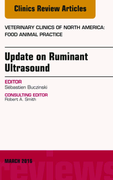
BOOK
Update on Ruminant Ultrasound, An Issue of Veterinary Clinics of North America: Food Animal Practice, E-Book
(2016)
Additional Information
Book Details
Abstract
This issue of Veterinary Clinics of North America: Food Animal Practice focuses on Ruminant Ultrasound. Article topics include: On-farm use of ultrasound for assessment of bovine respiratory disease, Echocardiography for the assessment of congenital heart defects in calves, Ultrasonography of the tympanic bulla and otitis media, Ultrasonography of the central nervous system and ultrasound guided CSF tap, Ultrasonographic examination of the abdomen of calves, Ascites in cattle: ultrasonographic findings and diagnosis, Ultrasonographic doppler use for reproduction management in heifers and cows, Ultrasound use for body condition and carcass quality assessment in cattle and lambs, and more!
Table of Contents
| Section Title | Page | Action | Price |
|---|---|---|---|
| Front Cover | Cover | ||
| Update on Ruminant Ultrasound\r | i | ||
| Copyright\r | ii | ||
| Contributors | iii | ||
| CONSULTING EDITOR | iii | ||
| EDITOR | iii | ||
| AUTHORS | iii | ||
| Contents | v | ||
| Preface: We Need More Studies on the Diagnostic and Prognostic Use of Ultrasound in Ruminants!\r | v | ||
| Specific Challenges in Conducting and Reporting Studies on the Diagnostic Accuracy of Ultrasonography in Bovine Medicine\r | v | ||
| On-Farm Use of Ultrasonography for Bovine Respiratory Disease\r | v | ||
| Echocardiography for the Assessment of Congenital Heart Defects in Calves\r | v | ||
| Ascites in Cattle: Ultrasonographic Findings and Diagnosis\r | vi | ||
| Ultrasonographic Examination of the Reticulum, Rumen, Omasum, Abomasum, and Liver in Calves\r | vi | ||
| Ultrasonographic Examination of the Reticulum, Rumen, Omasum, Abomasum,\rand Liver in Calves\r | vi | ||
| Ultrasonography of the Tympanic Bullae and Larynx in Cattle\r | vi | ||
| Ultrasound-Guided Nerve Block Anesthesia\r | vii | ||
| Ultrasonographic Doppler Use for Female Reproduction Management\r | vii | ||
| Methods for and Implementation of Pregnancy Diagnosis in Dairy Cows\r | vii | ||
| Practical Use of Ultrasound Scan in Small Ruminant Medicine and Surgery\r | vii | ||
| Ultrasound Use for Body Composition and Carcass Quality Assessment in Cattle and Lambs\r | viii | ||
| VETERINARY CLINICS OF\rNORTH AMERICA:\rFOOD ANIMAL PRACTICE\r | ix | ||
| FORTHCOMING ISSUES | ix | ||
| July 2016 | ix | ||
| November 2016 | ix | ||
| March 2017 | ix | ||
| RECENT ISSUES | ix | ||
| November 2015 | ix | ||
| July 2015 | ix | ||
| March 2015 | ix | ||
| Preface:\rWe Need More Studies on the\rDiagnostic and Prognostic Use of\rUltrasound in Ruminants!\r | xi | ||
| Specific Challenges in Conducting and Reporting Studies on the Diagnostic Accuracy of Ultrasonography in Bovine Medicine | 1 | ||
| Key points | 1 | ||
| INTRODUCTION | 1 | ||
| PHASES OF TESTING ASSESSMENT AND STUDY DESIGNS | 3 | ||
| REPORTING GUIDELINES FOR DIAGNOSTIC TEST ASSESSMENT STUDIES AS THEY RELATE TO IMAGING | 5 | ||
| VALIDITY AND BIASES IN DIAGNOSTIC TEST ASSESSMENT STUDIES AS THEY RELATE TO IMAGING USING ULTRASONOGRAPHY | 9 | ||
| SOURCES OF ERROR IN DIAGNOSTIC TEST ASSESSMENT STUDIES AS THEY RELATE TO IMAGING | 9 | ||
| INTERNAL VALIDATION OF ULTRASONOGRAPHY IN DIAGNOSTIC STUDIES: SYSTEMATIC ERROR | 11 | ||
| Biases That May Occur in Imaging Studies as a Result of the Approach to Patient Selection | 11 | ||
| Spectrum bias | 11 | ||
| Biases That May Occur in Imaging Studies as a Result of the Approach to Conducting the Index Test or Reference Text | 12 | ||
| Diagnostic review bias and incorporation bias | 12 | ||
| Classification bias | 12 | ||
| Biases in Imaging Studies as a Result of the Approach to Conducting the Flow of Patients Through the Study | 13 | ||
| Partial verification and differential verification | 13 | ||
| Loss to follow-up bias or indeterminate results | 13 | ||
| HOW TO PUT THE INCREMENTAL GAIN OF ULTRASONOGRAPHY OVER TRADITIONAL METHODS OF DIAGNOSIS TO PRACTICAL USE | 15 | ||
| SUPPLEMENTARY DATA | 17 | ||
| REFERENCES | 17 | ||
| On-Farm Use of Ultrasonography for Bovine Respiratory Disease | 19 | ||
| Key points | 19 | ||
| INTRODUCTION | 19 | ||
| ACCURACY OF THORACIC ULTRASONOGRAPHY IN YOUNG CATTLE | 20 | ||
| IMPLICATIONS OF ULTRASONOGRAPHIC LUNG LESIONS | 21 | ||
| ULTRASONOGRAPHIC EQUIPMENT | 22 | ||
| CALF PREPARATION AND RESTRAINT | 22 | ||
| APPROACH | 23 | ||
| THORACIC ULTRASONOGRAPHY TECHNIQUE | 24 | ||
| ULTRASONOGRAPHY SCORING | 27 | ||
| INDICATIONS AND IMPLEMENTATION | 32 | ||
| SUMMARY | 33 | ||
| REFERENCES | 33 | ||
| Echocardiography for the Assessment of Congenital Heart Defects in Calves | 37 | ||
| Key points | 37 | ||
| OVERVIEW | 37 | ||
| EQUIPMENT AND SETTINGS | 38 | ||
| PATIENT PREPARATION AND RESTRAINT | 38 | ||
| IMAGING APPROACH/PROTOCOL | 39 | ||
| Right Parasternal Views | 39 | ||
| Left Parasternal Views | 39 | ||
| Apical 4-Chamber/5-Chamber Views | 39 | ||
| SEQUENTIAL SEGMENTAL ANALYSIS | 39 | ||
| Step 1: Atrial Arrangement | 41 | ||
| Step 2: Ventricular Arrangement | 41 | ||
| Step 3: Atrioventricular Connections | 42 | ||
| Step 4: Morphology of the Atrioventricular Valves | 43 | ||
| Step 5: Ventriculoarterial Connections | 43 | ||
| Step 6: Morphology of the Arterial Valves | 43 | ||
| Step 7: Associated Malformations | 44 | ||
| SPECIFIC CONGENITAL MALFORMATIONS | 44 | ||
| Abnormal Communications (Shunts) | 44 | ||
| Atrial, atrioventricular, and ventricular septal defects | 44 | ||
| Patent ductus arteriosus | 47 | ||
| Aortopulmonary window | 47 | ||
| Outflow Tract Obstructions | 48 | ||
| Tetralogy/Pentalogy of Fallot | 51 | ||
| Coronary Abnormalities | 51 | ||
| Anomalies of Systemic and Pulmonary Venous Connections | 51 | ||
| Abnormalities of the Aorta and Aortic Arch | 51 | ||
| Abnormal Heart Location | 51 | ||
| HEMODYNAMIC CONSEQUENCES OF CONGENITAL HEART DISEASE | 51 | ||
| Assessment of the Hemodynamic Relevance of Ventricular Septal Defects | 52 | ||
| PROGNOSIS AND OUTCOMES OF CONGENITAL HEART DISEASE | 52 | ||
| SUPPLEMENTARY DATA | 53 | ||
| REFERENCES | 53 | ||
| Ascites in Cattle | 55 | ||
| Key points | 55 | ||
| INTRODUCTION | 55 | ||
| DIAGNOSTIC PROCEDURE IN SUSPECTED CASES OF ASCITES | 56 | ||
| Clinical Examination | 56 | ||
| Blood Examination | 56 | ||
| Ultrasonographic Examination | 56 | ||
| Abdominocentesis | 56 | ||
| TECHNIQUES OF ABDOMINAL ULTRASONOGRAPHY AND ABDOMINOCENTESIS | 56 | ||
| Ultrasonography Technique in Suspected Cases of Ascites | 56 | ||
| Technique for Abdominocentesis in Suspected Cases of Ascites | 57 | ||
| TYPES OF FREE PERITONEAL FLUID | 58 | ||
| NONINFLAMMATORY ASCITES | 58 | ||
| Clinical Signs of Noninflammatory Ascites | 59 | ||
| Ultrasonographic Findings of Noninflammatory Ascites | 60 | ||
| Causes of Noninflammatory Ascites | 61 | ||
| Right-Sided Cardiac Insufficiency as the Cause of Ascites | 62 | ||
| Mediastinal Masses as the Cause of Ascites | 62 | ||
| Liver Disease as the Cause of Ascites | 63 | ||
| Kidney Disease as the Cause of Ascites | 65 | ||
| Ileus as the Cause of Ascites | 65 | ||
| Tumors of the Peritoneum, Serous Membranes, and Omentum | 65 | ||
| Obstruction and Compression of the Caudal Vena Cava as the Cause of Ascites | 66 | ||
| INFLAMMATORY ASCITES (PERITONITIS) | 68 | ||
| Ultrasonographic Findings of Focal and Generalized Peritonitis | 70 | ||
| Omental Bursitis | 75 | ||
| UROPERITONEUM | 77 | ||
| HEMOPERITONEUM | 78 | ||
| CHYLOUS ASCITES | 78 | ||
| BILIARY ASCITES (BILE PERITONITIS) | 79 | ||
| DIFFERENTIAL DIAGNOSIS OF INTRA-ABDOMINAL FLUID ACCUMULATION | 79 | ||
| REFERENCES | 80 | ||
| Ultrasonographic Examination of the Reticulum, Rumen, Omasum, Abomasum, and Liver in Calves | 85 | ||
| Key points | 85 | ||
| INTRODUCTION | 85 | ||
| TECHNIQUE OF ULTRASONOGRAPHIC EXAMINATION OF CALVES | 86 | ||
| RETICULUM | 86 | ||
| Examination of the Reticulum | 86 | ||
| Ultrasonographic Findings of the Reticulum | 86 | ||
| RUMEN | 87 | ||
| Examination of the Rumen | 87 | ||
| Ultrasonographic Findings of the Rumen | 88 | ||
| OMASUM | 90 | ||
| Examination of the Omasum | 90 | ||
| Ultrasonographic Findings of the Omasum | 90 | ||
| ABOMASUM | 91 | ||
| Examination of the Abomasum | 91 | ||
| Ultrasonographic Findings of the Abomasum | 92 | ||
| ESOPHAGEAL GROOVE REFLEX AND FACTORS AFFECTING GROOVE CLOSURE | 98 | ||
| Ultrasonographic Monitoring of the Esophageal Groove Reflex | 98 | ||
| Examination of the Esophageal Groove Reflex | 98 | ||
| RUMINAL DRINKER SYNDROME | 98 | ||
| LIVER | 100 | ||
| Ultrasonographic Examination of the Liver | 100 | ||
| Ultrasonographic Findings of the Liver and Gallbladder | 101 | ||
| Abnormal Ultrasonographic Findings of the Liver and Gallbladder | 105 | ||
| UMBILICUS | 105 | ||
| SUMMARY | 105 | ||
| REFERENCES | 106 | ||
| Ultrasonographic\rExamination of the Spinal\rCord and Collection of\rCerebrospinal Fluid from the\rAtlanto-Occipital Space in Cattle\r | 109 | ||
| Key points | 109 | ||
| INTRODUCTION | 109 | ||
| ANATOMY OF THE ATLANTO-OCCIPITAL SPACE | 110 | ||
| ULTRASONOGRAPHIC EXAMINATION OF THE SPINAL CORD FROM THE ATLANTO-OCCIPITAL SPACE | 111 | ||
| Preparation of Cattle for the Ultrasonographic Examination | 111 | ||
| Technique of Ultrasonographic Examination | 111 | ||
| Ultrasonographic Findings of the Atlanto-Occipital Space | 111 | ||
| Measurements in the Atlanto-Occipital Space in 73 Euthanized Cows | 112 | ||
| ULTRASOUND-GUIDED COLLECTION OF CEREBROSPINAL FLUID FROM THE ATLANTO-OCCIPITAL SPACE | 114 | ||
| Preparation of Cattle and Cerebrospinal Fluid Collection Technique | 114 | ||
| Examination of the Cerebrospinal Fluid | 115 | ||
| ULTRASONOGRAPHIC EXAMINATION OF THE SPINAL CORD FROM THE LUMBOSACRAL AREA IN THE CALF | 116 | ||
| SUMMARY | 117 | ||
| REFERENCES | 117 | ||
| Ultrasonography of the Tympanic Bullae and Larynx in Cattle | 119 | ||
| Key points | 119 | ||
| INTRODUCTION | 119 | ||
| ULTRASONOGRAPHY OF THE TYMPANIC BULLA | 120 | ||
| Anatomy of the Tympanic Bulla | 120 | ||
| Technique | 120 | ||
| Contention and preparation | 121 | ||
| Positioning of the probe | 121 | ||
| Transverse position | 122 | ||
| Longitudinal position | 122 | ||
| Normal Images | 122 | ||
| Abnormal Images | 124 | ||
| Diagnostic Value | 124 | ||
| ULTRASONOGRAPHY OF THE LARYNX | 126 | ||
| Anatomy of the Larynx | 126 | ||
| Technique and Normal Images | 127 | ||
| Abnormal Images and Diagnostic Value | 130 | ||
| ACKNOWLEDGMENTS | 130 | ||
| REFERENCES | 130 | ||
| Ultrasound-Guided Nerve Block Anesthesia | 133 | ||
| Key points | 133 | ||
| INTRODUCTION | 133 | ||
| ULTRASOUND IN REGIONAL ANESTHESIA | 134 | ||
| ADVANTAGES OF ULTRASOUND GUIDANCE IN REGIONAL ANESTHESIA | 134 | ||
| Visualization of Anatomic Structures | 134 | ||
| Reduction of Local Anesthetic Requirements | 135 | ||
| Improvement in Anesthetic Block Quality | 135 | ||
| Prevention of Fatalities | 135 | ||
| LOCALIZATION OF NERVES OF CLINICAL RELEVANCE | 135 | ||
| Anatomy and Ultrasound Image of the Nerve | 135 | ||
| Needle Visualization and Guidance | 136 | ||
| The Doughnut Sign | 136 | ||
| PERINEURAL BLOCK TECHNIQUES | 137 | ||
| Paravertebral Nerve Block | 137 | ||
| Description | 137 | ||
| Technique | 138 | ||
| Caudal Epidural Block | 139 | ||
| Ultrasonographic Doppler Use for Female Reproduction Management | 149 | ||
| Key points | 149 | ||
| INTRODUCTION | 149 | ||
| PRINCIPLE OF DOPPLER ULTRASONOGRAPHY | 150 | ||
| TECHNIQUE OF DOPPLER ULTRASONOGRAPHY | 150 | ||
| Evaluation of Blood Flow | 150 | ||
| Transrectal Localization of the Uterine Artery | 152 | ||
| UTERINE BLOOD FLOW | 153 | ||
| Estrous Cycle | 153 | ||
| Pregnancy | 154 | ||
| Puerperium | 156 | ||
| OVARIAN BLOOD FLOW | 157 | ||
| Follicular Blood Flow | 157 | ||
| Luteal Blood Flow | 158 | ||
| SUMMARY | 160 | ||
| REFERENCES | 161 | ||
| Methods for and Implementation of Pregnancy Diagnosis in Dairy Cows | 165 | ||
| Key points | 165 | ||
| ATTRIBUTES OF THE IDEAL PREGNANCY TEST | 166 | ||
| RETURN TO ESTRUS AS A DIAGNOSTIC INDICATOR OF PREGNANCY STATUS | 166 | ||
| PREGNANCY LOSS IN LACTATING DAIRY COWS | 166 | ||
| DIRECT METHODS FOR PREGNANCY DIAGNOSIS | 168 | ||
| Transrectal Palpation | 168 | ||
| B-Mode Ultrasonography | 168 | ||
| Problems with Early Pregnancy Diagnosis Using Transrectal Ultrasonography | 169 | ||
| INDIRECT METHODS FOR PREGNANCY DIAGNOSIS IN DAIRY COWS | 171 | ||
| Progesterone | 171 | ||
| Pregnancy-Associated Proteins | 173 | ||
| Pregnancy-Associated Glycoproteins | 173 | ||
| FUTURE TECHNOLOGIES FOR PREGNANCY DIAGNOSIS | 176 | ||
| SUMMARY | 177 | ||
| REFERENCES | 177 | ||
| Practical Use of Ultrasound Scan in Small Ruminant Medicine and Surgery | 181 | ||
| Key points | 181 | ||
| INTRODUCTION | 181 | ||
| ULTRASONOGRAPHIC EXAMINATION—EQUIPMENT | 182 | ||
| SITES FOR ULTRASONOGRAPHIC EXAMINATION | 182 | ||
| Chest | 182 | ||
| Liver | 182 | ||
| Bladder, Uterus, Vagina, and Ventral Abdomen | 182 | ||
| Right Kidney | 183 | ||
| Abomasal Diameter of Neonatal Lambs | 183 | ||
| Vaginal Prolapse | 183 | ||
| Scrotum | 183 | ||
| Joints | 183 | ||
| ULTRASONOGRAPHIC FINDINGS | 183 | ||
| Chest | 183 | ||
| Lung Consolidation | 183 | ||
| Pleural Effusion | 185 | ||
| Pleural/Lung Abscesses | 185 | ||
| Fibrinous Pleurisy | 186 | ||
| Ovine Pulmonary Adenocarcinoma | 186 | ||
| HEART | 189 | ||
| Congenital Cardiac Defects | 189 | ||
| Vegetative Endocarditis | 190 | ||
| Pericarditis | 190 | ||
| ABDOMEN | 190 | ||
| Ascites | 190 | ||
| Peritonitis | 191 | ||
| Small Intestinal Torsion | 192 | ||
| Intra-abdominal Hemorrhage | 192 | ||
| Liver | 192 | ||
| Liver Fluke | 193 | ||
| URINARY TRACT | 194 | ||
| Kidney | 195 | ||
| SCROTUM | 196 | ||
| Testicular Atrophy | 196 | ||
| Epididymitis | 197 | ||
| PREGNANCY | 198 | ||
| Obstetric Problems | 198 | ||
| Uterine Torsion | 199 | ||
| VAGINAL PROLAPSE | 200 | ||
| UDDER | 200 | ||
| JOINTS | 201 | ||
| MUSCLE | 201 | ||
| ABOMASUM IN NEONATES | 202 | ||
| BRAIN | 202 | ||
| DISCUSSION | 202 | ||
| SUPPLEMENTARY DATA | 203 | ||
| REFERENCES | 203 | ||
| Ultrasound Use for Body Composition and Carcass Quality Assessment in Cattle and Lambs | 207 | ||
| Key points | 207 | ||
| INTRODUCTION | 207 | ||
| INDICATIONS/CONTRAINDICATIONS | 208 | ||
| Certification Requirements (Equipment and Technician) | 209 | ||
| Expectations of Producer/Prescanning Management of the Animal | 210 | ||
| Application of Ultrasound for Carcass Traits in Beef Feedlot Settings | 210 | ||
| Lamb Industry to Beef Industry Comparison on the Use of Ultrasound for Body Composition Traits | 211 | ||
| TECHNIQUE/PROCEDURE | 211 | ||
| Patient Positioning and Preparation | 211 | ||
| Approach | 212 | ||
| Specific Technique/Procedure | 212 | ||
| COMPLICATIONS AND MANAGEMENT | 214 | ||
| POSTOPERATIVE CARE | 215 | ||
| REPORTING, FOLLOW-UP, AND CLINICAL IMPLICATIONS | 216 | ||
| OUTCOMES | 216 | ||
| CURRENT CONTROVERSIES | 216 | ||
| FUTURE CONSIDERATIONS | 216 | ||
| SUMMARY | 217 | ||
| REFERENCES | 217 | ||
| Index | 219 |
