Additional Information
Book Details
Abstract
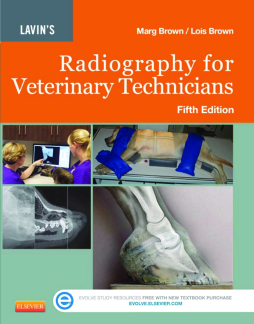
Written by veterinary technicians for veterinary students and practicing technicians, Lavin’s Radiography for Veterinary Technicians, 5th Edition, combines all the aspects of imaging — including production, positioning, and evaluation of radiographs —into one comprehensive text. Completely updated with all new vivid, color equipment photos, positioning drawings and detailed anatomy drawings, this fifth edition is a valuable resource for students, technicians and veterinarians who need information on the latest technology or unique positioning.
- Broad coverage of radiologic science, physics, imaging and protection provide you with foundations for good technique.
- Positioning photos, radiographic images and anatomical drawings presented side-by-side with text explanation for each procedure increases your comprehension and retention.
- Objectives, key terms, outlines, chapter introductions and key points help you organize information to ensure you understand what is most important in every chapter.
- NEW! More than 1000 new full-color photos and updated radiographic images visually demonstrate the relationship between anatomy and positioning.
- NEW! All-new color anatomy art created by an expert medical illustrator help you to recognize and avoid making imaging mistakes.
- NEW! Non-Manual restraint techniques including sandbags, tape, rope, sponges, sedation and combinations improve your safety and radiation protection.
- NEW! Chapter on dental radiography aids general veterinarian techs and those specializing in dentistry.
- NEW! Increased emphasis on digital radiography, including quality factors and post-processing, keeps you up-to-date on the most recent developments in digital technology.
Table of Contents
| Section Title | Page | Action | Price |
|---|---|---|---|
| Front cover | cover | ||
| Half title page | i | ||
| Evolve page | ii | ||
| Lavin's Radiography for Veterinary Technicians, 5/e | iii | ||
| Copyright page | iv | ||
| Contributors | v | ||
| Dedication | vi | ||
| Preface | vii | ||
| Acknowledgments | ix | ||
| Credits | xi | ||
| Table of Contents | xv | ||
| One Diagnostic Imaging | 1 | ||
| One The Technical Side of Imaging | 2 | ||
| 1 Basic Concepts | 2 | ||
| Outline | 2 | ||
| Learning Objectives | 2 | ||
| Key Terms | 2 | ||
| Arithmetic | 3 | ||
| Fractions | 3 | ||
| Addition and Subtraction | 3 | ||
| Whole Numbers | 3 | ||
| Proportionality (Variation) of Numbers | 3 | ||
| Direct Proportionality | 3 | ||
| Indirect Proportionality | 3 | ||
| Units of Measurement | 3 | ||
| Mass | 3 | ||
| Length | 3 | ||
| Time | 3 | ||
| Prefixes | 4 | ||
| Summary | 4 | ||
| Review Questions | 4.e1 | ||
| 2 The Atom and Radioactivity | 5 | ||
| Outline | 5 | ||
| Learning Objectives | 5 | ||
| Key Terms | 5 | ||
| The Elements | 6 | ||
| Atomic Theory | 6 | ||
| Atomic Structure | 6 | ||
| The Electron | 6 | ||
| The Proton | 8 | ||
| The Neutron | 9 | ||
| Atomic Weight | 9 | ||
| Atomic Nomenclature | 9 | ||
| Atomic Representation | 9 | ||
| Combinations of Atoms | 9 | ||
| The Orderly Organization of Matter | 9 | ||
| Isotopes | 9 | ||
| Radioactivity | 9 | ||
| Ionization | 9 | ||
| Summary | 10 | ||
| Review Questions | 10.e1 | ||
| 3 Electrostatics and Energy, Magnetism and Electricity | 11 | ||
| Outline | 11 | ||
| Learning Objectives | 11 | ||
| Key Terms | 11 | ||
| Electrostatics and Energy | 12 | ||
| Matter | 12 | ||
| Energy | 12 | ||
| Electrical Energy | 12 | ||
| Electromagnetic Spectrum | 12 | ||
| Particle-Wave Theory | 12 | ||
| Frequency | 12 | ||
| Energy in Radiography | 14 | ||
| Properties of X-Rays | 14 | ||
| Rœntgen’s Properties of Rays | 14 | ||
| Summary | 14 | ||
| Magnetism and Electricity | 15 | ||
| Electricity Becomes X-Rays | 15 | ||
| Electrostatics | 15 | ||
| Laws of Electrostatics | 15 | ||
| Electrification | 16 | ||
| Contact | 16 | ||
| Friction | 16 | ||
| Induction | 17 | ||
| Conductors and Insulators | 17 | ||
| Electric Current | 17 | ||
| Resistance | 17 | ||
| Potential Difference | 17 | ||
| Voltage | 18 | ||
| Summary | 18 | ||
| Review Questions | 18.e1 | ||
| Electrostatics and Energy | 18.e1 | ||
| Magnetism and Electricity | 18.e1 | ||
| 4 Diagnostic X-Ray Production | 19 | ||
| Outline | 19 | ||
| Learning Objectives | 19 | ||
| Key Terms | 19 | ||
| Diagnostic X-Ray Production | 20 | ||
| Power | 20 | ||
| The Electrical Circuit | 20 | ||
| The X-Ray Circuit Is Actually a Circle | 20 | ||
| On/Off Switch | 20 | ||
| Wall Switch | 20 | ||
| Line Voltage Compensator | 21 | ||
| Circuit Breakers: Amperage and Ground | 22 | ||
| Amperage | 22 | ||
| Ground | 22 | ||
| Direct Current and Alternating Current | 22 | ||
| Transformers | 23 | ||
| Rectifiers | 24 | ||
| Three-Phase Circuits | 25 | ||
| High Frequency | 25 | ||
| The Outside of the X-Ray Unit | 26 | ||
| Large Animal Portable X-Ray Units | 27 | ||
| Diagnostic X-Ray Production | 27 | ||
| The X-Ray Tube | 27 | ||
| The Cathode | 28 | ||
| The Anode | 30 | ||
| The Rotating Anode | 30 | ||
| The Rotor Circuit | 30 | ||
| The Stationary Anode | 31 | ||
| The Line Focus Principle | 31 | ||
| Off-Focus Radiation and Heat Bloom | 31 | ||
| Heat Dissipation | 31 | ||
| The Tube Rating Chart | 32 | ||
| Focal Spot Bloom | 33 | ||
| The Anode Heel Effect | 33 | ||
| The Exposure Switch | 34 | ||
| Exposure Switch Variations | 35 | ||
| Summary | 35 | ||
| Review Questions | 35.e1 | ||
| Two The Application of X-rays and the Presentation of the Image | 36 | ||
| 5 Imaging on Film* | 36 | ||
| Outline | 36 | ||
| Learning Objectives | 36 | ||
| Key Terms | 36 | ||
| A Brief History of Terminology | 37 | ||
| Film-Based Imaging | 38 | ||
| X-Ray Cassettes | 38 | ||
| Film/Screen Contact | 39 | ||
| Intensifying Screens | 42 | ||
| Manufacture | 42 | ||
| Characteristics | 42 | ||
| The Blue/Green Question | 44 | ||
| More about Color | 44 | ||
| Screen Speed | 45 | ||
| Luminescence | 45 | ||
| Screen Aging Response | 46 | ||
| Screen Artifacts | 48 | ||
| Chemistry Spills | 48 | ||
| Screen Cleaners | 49 | ||
| Radiography Film | 49 | ||
| World’s First Human Portrait | 50 | ||
| Composition of Radiography Film | 51 | ||
| Film Base | 51 | ||
| Film Emulsion | 52 | ||
| The Supercoat | 53 | ||
| Latent Image Formation | 53 | ||
| Base + Fog | 54 | ||
| Film Response to Light | 54 | ||
| Film Colors | 54 | ||
| Film Speed | 55 | ||
| Film Speed and White Box Film | 55 | ||
| Resolution | 55 | ||
| Storage: Cassettes, Screens, and Films | 56 | ||
| Film | 56 | ||
| Cassettes and Screens | 57 | ||
| Summary | 57 | ||
| Review Questions | 57.e1 | ||
| 6 Producing the Image | 58 | ||
| Outline | 58 | ||
| Learning Objectives | 58 | ||
| Key Terms | 58 | ||
| The Radiography Unit in Use | 59 | ||
| Film Fog | 59 | ||
| The Technical Factors | 61 | ||
| Radiographic Contrast versus Subject Contrast | 61 | ||
| Distance, Kilovoltage, Milliamperage, Time | 61 | ||
| Distance and the Inverse Square Law | 61 | ||
| Kilovoltage, Milliamperage, and Time | 62 | ||
| Optimizing Kilovoltage | 63 | ||
| The 15% Rule | 64 | ||
| Milliamperage | 64 | ||
| Adjusting the Factors | 65 | ||
| Developing Technique Charts | 65 | ||
| The Three “Commandments” | 65 | ||
| Two Radiographic Positioning and Related Anatomy | 173 | ||
| 17 Overview of Positioning | 174 | ||
| Outline | 174 | ||
| Learning Objectives | 174 | ||
| Key Terms | 174 | ||
| Positional Terminology | 175 | ||
| Rules of Positioning | 175 | ||
| Limb Terminology | 175 | ||
| Patient Positioning | 176 | ||
| The Patient | 176 | ||
| Patient Preparation | 177 | ||
| Human Safety | 178 | ||
| Positioning Aids | 178 | ||
| Required Views and Positioning Guidelines | 179 | ||
| Image Identification | 181 | ||
| Viewing Radiographs | 182 | ||
| Radiographic Checklist | 182 | ||
| Review Questions | 183.e1 | ||
| Bibliography | 183 | ||
| 18 Small Animal Abdomen | 184 | ||
| Outline | 184 | ||
| Learning Objectives | 184 | ||
| Key Terms | 184 | ||
| Radiographic Concerns | 185 | ||
| Positions | 186 | ||
| Lateral | 186 | ||
| Measure: | 186 | ||
| Central Ray: | 186 | ||
| Canine: | 186 | ||
| Feline: | 186 | ||
| Borders: | 186 | ||
| Positioning | 186 | ||
| Comments and Tips | 186 | ||
| Ventrodorsal | 188 | ||
| Measure: | 188 | ||
| Central Ray: | 188 | ||
| Canine: | 188 | ||
| Feline: | 188 | ||
| Borders: | 188 | ||
| Positioning | 188 | ||
| Comments and Tips | 189 | ||
| Further Views | 190 | ||
| Dorsoventral | 190 | ||
| Measure, Central Ray, Borders: | 190 | ||
| Positioning | 190 | ||
| Comments and Tips | 190 | ||
| Lateral Decubitus (Ventrodorsal View with Horizontal Beam) | 191 | ||
| Measure, Central Ray, Borders: | 191 | ||
| Positioning | 191 | ||
| Comments and Tips | 191 | ||
| Modified Lateral and Lateral Oblique | 192 | ||
| Both Positions | 192 | ||
| Measure: | 192 | ||
| Central Ray: | 192 | ||
| Borders: | 192 | ||
| Modified Lateral | 192 | ||
| Glossary | 513 | ||
| Index | 525 | ||
| A | 525 | ||
| B | 526 | ||
| C | 526 | ||
| D | 528 | ||
| E | 530 | ||
| F | 530 | ||
| G | 532 | ||
| H | 532 | ||
| I | 533 | ||
| J | 534 | ||
| K | 534 | ||
| L | 534 | ||
| M | 535 | ||
| N | 536 | ||
| O | 537 | ||
| P | 537 | ||
| Q | 538 | ||
| R | 538 | ||
| S | 539 | ||
| T | 542 | ||
| U | 543 | ||
| V | 543 | ||
| W | 544 | ||
| X | 544 |
