Additional Information
Book Details
Abstract
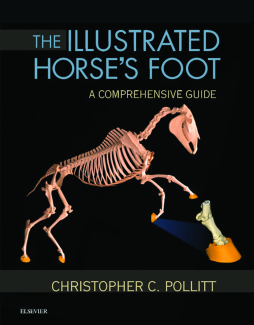
Achieve optimal results in equine foot care and treatment! The Illustrated Horse’s Foot: A Comprehensive Guide uses clear instructions in an atlas-style format to help you accurately identify, diagnose, and treat foot problems in horses. Full-color clinical photographs show structure and function as well as the principles of correct clinical examination and shoeing, and a companion website has videos depicting equine foot cases. Written by internationally renowned expert Christoher Pollitt, this resource enhances your ability to treat equine conditions ranging from laminitis to foot cracks, infections, trauma, vascular compromise, and arthritis.
- Comprehensive coverage addresses a wide range of equine foot conditions.
- A unique collection of MIMICs provides beautifully detailed anatomical hoof images.
- 284 high-quality images show conditions of the equine foot, including many 2-D reconstructions of MRI and CT data.
- Step-by-step case histories follow equine patients from initial presentation through diagnosis to treatment and outcome.
- A convenient, templated format provides quick access to clinical signs, diagnosis, treatment, and prognosis.
- Expert author Chris Pollitt is a pioneer in the use of advanced radiographic, CT, and MRI technology for imaging equine foot and laminitis problems to facilitate accurate diagnosis and effective treatment.
- A companion website located at pollitthorsesfoot.com located at pollitthorsesfoot.com includes video clips of equine foot cases.
Table of Contents
| Section Title | Page | Action | Price |
|---|---|---|---|
| Front cover | Cover | ||
| The illustrated horse’s foot | 1 | ||
| Copyright | ii | ||
| Dedication | iii | ||
| Preface | iv | ||
| Acknowledgments | vi | ||
| Introduction | vii | ||
| References | vii | ||
| Table of contents | viii | ||
| 1 Foot Structure and Function | 1 | ||
| 1 The hoof | 3 | ||
| The hoof wall | 3 | ||
| The stratum externum (periople) | 3 | ||
| The stratum medium (coronary horn) | 4 | ||
| The stratum internum (epidermal lamellae) | 4 | ||
| Secondary epidermal lamellae | 4 | ||
| The basement membrane | 4 | ||
| Hemidesmosomes | 4 | ||
| Basal cell cytoskeleton | 5 | ||
| Hoof wall growth | 5 | ||
| The dermis | 5 | ||
| The coronary dermis | 6 | ||
| The sole dermis | 6 | ||
| References | 6 | ||
| 2 The hoof capsule | 7 | ||
| The tubular hoof wall | 7 | ||
| The dermis | 8 | ||
| The coronary dermis | 8 | ||
| References | 20 | ||
| 3 The dermis | 21 | ||
| The coronary dermis | 21 | ||
| The sole corium | 21 | ||
| References | 29 | ||
| 4 The mimics anatomic models | 30 | ||
| 5 Planar anatomy | 38 | ||
| Anatomic terms | 38 | ||
| Planar anatomy | 38 | ||
| Sagittal and parasagittal anatomy | 39 | ||
| Gross anatomy from sections | 39 | ||
| References | 58 | ||
| 6 The suspensory apparatus of the distal phalanx | 59 | ||
| References | 71 | ||
| 7 The circulatory system | 72 | ||
| The terminal arch—the source | 73 | ||
| The arterial system (forelimb): A classical description | 73 | ||
| The arterial system of the hindlimb: A classic description | 74 | ||
| The venous system of the foot | 75 | ||
| The microcirculation | 76 | ||
| Components of the microcirculation | 76 | ||
| Delivery of glucose and exchange of water and other materials occur across capillaries | 77 | ||
| Molecules pass between and through capillary endothelial cells | 77 | ||
| Diffusion of molecules or particles | 77 | ||
| Equilibrium in the capillary bed | 77 | ||
| Venules collect blood from capillaries and act as a blood reservoir | 77 | ||
| Arteriovenous anastomoses | 77 | ||
| References | 124 | ||
| 8 Histology | 125 | ||
| Lamellar histology | 125 | ||
| Transverse histology | 127 | ||
| Dorsal histology | 140 | ||
| References | 145 | ||
| 9 Electron microscopy | 146 | ||
| 10 Bones | 152 | ||
| 11 Joints | 162 | ||
| Distal interphalangeal joint, or coffin joint | 162 | ||
| Metacarpophalangeal, or fetlock joint | 162 | ||
| References | 167 | ||
| 12 Radiography of the foot | 168 | ||
| Radiographic technique | 168 | ||
| Venography | 168 | ||
| Venographic technique | 168 | ||
| Digital radiography and venograms | 169 | ||
| References | 173 | ||
| 13 The palmar digital nerve | 174 | ||
| References | 174 | ||
| 2 Conditions of the Foot | 175 | ||
| 14 Laminitis | 177 | ||
| Laminitis radiology | 177 | ||
| Radiology of acute/early chronic laminitis | 177 | ||
| The rate at which the hdpd increases correlates with the severity of the acute lesion | 177 | ||
| Radiology of chronic laminitis | 178 | ||
| Venography of laminitis | 179 | ||
| Laminitis histopathology | 180 | ||
| Grade 0: Normal lamellar histopathology | 180 | ||
| Grade 1: Laminitis histopathology | 180 | ||
| Grade 2: Laminitis histopathology | 180 | ||
| Grade 3: Laminitis histopathology | 180 | ||
| Case history 1: Peracute, severe laminitis in an arabian mare | 188 | ||
| Case history 2: Severe chroinic laminitis in a thoroughbred colt | 194 | ||
| Insulin and laminitis | 201 | ||
| Equine metabolic syndrome | 201 | ||
| Hyperinsulinemic laminitis | 201 | ||
| Case history 3: Hyperinsulinemic laminitis in aged pony | 201 | ||
| Case history 4: Traumatic laminitis (road founder) in a seven-year-old thoroughbred gelding | 210 | ||
| Concluding remarks | 216 | ||
| Case history 5: Chronic laminitis in an arabian endurance horse | 216 | ||
| References | 221 | ||
| 15 Navicular disease | 223 | ||
| References | 228 | ||
| 16 Midline toe cracks | 229 | ||
| Case history 1: Full-length toe cracks in the forefeet of a mature thoroughbred mare | 229 | ||
| Case history 2: Distal toe cracks in the forefeet of a thoroughbred broodmare | 234 | ||
| Case history 3: Full length toe cracks in the forefeet of a quarterhorse stallion | 235 | ||
| Case history 4: Full length toe cracks in the forefeet of a thoroughbred filly | 239 | ||
| 17 Seedy toe (white line disease) | 241 | ||
| 18 Ossification of the ungular cartilages | 248 | ||
| References | 250 | ||
| 19 Coronary band injury | 251 | ||
| Coronary band stake wounds | 251 | ||
| Case history 1: Coronary band stake wound to the foot of an endurance horse | 251 | ||
| Case history 2: Coronary band stake wound to the foot of a trail riding pony | 251 | ||
| 20 Infected nail holes | 254 | ||
| Case history 1: Infected nail hole with coronet abscess | 255 | ||
| Case history 2: Infected nail hole in the foot of a competition horse | 255 | ||
| Index | 261 | ||
| A | 261 | ||
| B | 261 | ||
| C | 261 | ||
| D | 261 | ||
| E | 262 | ||
| F | 262 | ||
| G | 262 | ||
| H | 262 | ||
| I | 262 | ||
| J | 262 | ||
| L | 262 | ||
| M | 263 | ||
| N | 263 | ||
| O | 263 | ||
| P | 263 | ||
| Q | 263 | ||
| R | 263 | ||
| S | 264 | ||
| T | 264 | ||
| U | 264 | ||
| V | 264 | ||
| W | 264 | ||
| End sheet | ES4 |
