Additional Information
Book Details
Abstract
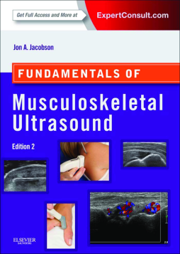
FUNDAMENTALS OF MUSCULOSKELETAL ULTRASOUND packs a big punch for such a compact book. It teaches the resident, clinician and even medical student, how to perform and read musculoskeletal ultrasounds, while highlighting the basic anatomy needed to perform and interpret ultrasounds and the salient points needed to make diagnosis. Key anatomy, concepts, diseases and even controversies are highlighted, rather than presenting a lengthy tome covering the A to Z's of musculoskeletal ultrasound.
- Organized in a simple, outline format (emphasizing lists and tables) for easy access to information.
- Features almost 1200 high quality images that clearly demonstrate essential concepts, techniques and interpretation skills.
- Provides step-by-step instructions on how to perform musculoskeletal ultrasound techniques and interpret musculoskeletal ultrasound findings.
- Reviews sonographic anatomy of peripheral joints to help you understand the anatomy so you can interpret ultrasound scans with confidence.
- Reviews the sonographic appearances of common musculoskeletal pathologies to clearly differentiate one condition from another.
Table of Contents
| Section Title | Page | Action | Price |
|---|---|---|---|
| Front Cover | cover | ||
| Inside Front Cover | ifc1 | ||
| Fundamentals of Musculoskeletal Ultrasound, 2/e | i | ||
| Copyright Page | iv | ||
| Dedication | v | ||
| Preface | vii | ||
| Acknowledgments | ix | ||
| Table Of Contents | xi | ||
| Cine Clip Video Contents | xiii | ||
| 1 Introduction | xiii | ||
| 2 Basic Pathology Concepts | xiii | ||
| 3 Shoulder Ultrasound | xiii | ||
| 4 Elbow Ultrasound | xiv | ||
| 5 Wrist and Hand Ultrasound | xiv | ||
| 6 Hip and Thigh Ultrasound | xv | ||
| 7 Knee Ultrasound | xv | ||
| 8 Ankle, Foot, and Lower Leg Ultrasound | xv | ||
| 9 Interventional Techniques | xvi | ||
| 1 Introduction | 1 | ||
| Chapter Outline | 1 | ||
| Equipment Considerations and Image Formation | 1.e1 | ||
| Scanning Technique | 1.e2 | ||
| Image Appearance | 1.e3 | ||
| Sonographic Appearances of Normal Structures | 1.e5 | ||
| Sonographic Artifacts | 1.e6 | ||
| Miscellaneous Ultrasound Techniques | 1.e10 | ||
| Color and Power Doppler | 1.e12 | ||
| Dynamic Imaging | 1.e12 | ||
| References | 1.e14 | ||
| 2 Basic Pathology Concepts | 2 | ||
| Chapter Outline | 2 | ||
| Muscle and Tendon Injury | 2.e1 | ||
| Bone Injury | 2.e6 | ||
| Infection | 2.e8 | ||
| Arthritis | 2.e12 | ||
| Rheumatoid Arthritis | 2.e12 | ||
| Psoriatic Arthritis | 2.e14 | ||
| Gout | 2.e15 | ||
| Osteoarthritis | 2.e15 | ||
| Myositis and Diabetic Muscle Infarction | 2.e17 | ||
| Soft Tissue Foreign Bodies | 2.e17 | ||
| Peripheral Nerve Entrapment | 2.e21 | ||
| Soft Tissue Masses | 2.e23 | ||
| Lipoma | 2.e23 | ||
| Peripheral Nerve Sheath Tumors | 2.e25 | ||
| Vascular Anomalies | 2.e26 | ||
| Ganglion Cysts | 2.e28 | ||
| Lymph Nodes | 2.e29 | ||
| Malignant Soft Tissue Tumors | 2.e29 | ||
| Bone Masses | 2.e33 | ||
| References | 2.e37 | ||
| 3 Shoulder Ultrasound | 3 | ||
| Chapter Outline | 3 | ||
| Ultrasound Examination Technique | 5 | ||
| General Comments | 5 | ||
| Position No. 1: Long Head of Biceps Brachii Tendon | 5 | ||
| Position No. 2: Subscapularis and Biceps Tendon Dislocation | 7 | ||
| Position No. 3: Supraspinatus and Infraspinatus | 8 | ||
| Position No. 4: Acromioclavicular Joint, Subacromial-Subdeltoid Bursa, and Dynamic Evaluation | 15 | ||
| Position No. 5: Infraspinatus, Teres Minor, and Posterior Glenoid Labrum | 16 | ||
| Rotator Cuff Abnormalities | 18 | ||
| Supraspinatus Tears and Tendinosis | 18 | ||
| General Comments | 18 | ||
| Partial-Thickness Tear | 20 | ||
| Full-Thickness Tear | 25 | ||
| Tendinosis | 29 | ||
| Indirect Signs of Supraspinatus Tendon Tear | 30 | ||
| Tendon Thinning. | 30 | ||
| Cortical Irregularity. | 31 | ||
| Joint Effusion and Bursal Fluid. | 31 | ||
| Cartilage Interface Sign. | 32 | ||
| Infraspinatus Tears and Tendinosis | 32 | ||
| Subscapularis Tears and Tendinosis | 33 | ||
| Rotator Cuff Atrophy | 33 | ||
| Postoperative Shoulder | 36 | ||
| Calcific Tendinosis | 38 | ||
| Impingement Syndrome | 40 | ||
| Adhesive Capsulitis | 42 | ||
| Pitfalls in Rotator Cuff Ultrasound | 44 | ||
| Errors in Scanning Technique | 44 | ||
| Improper Positioning of the Shoulder | 44 | ||
| Incomplete Evaluation of the Supraspinatus Tendon | 44 | ||
| Imaging of the Rotator Cuff Too Distally | 44 | ||
| Misinterpretation of Normal Structures | 45 | ||
| Misinterpretation of the Rotator Interval | 45 | ||
| Misinterpretation of the Musculotendinous Junction | 46 | ||
| Misinterpretation of the Supraspinatus-Infraspinatus Junction | 46 | ||
| Misinterpretation of Pathology | 47 | ||
| Subacromial-Subdeltoid Bursa Simulating Tendon | 47 | ||
| Rim-Rent Tear Versus Intrasubstance Tear | 48 | ||
| Tendinosis Versus Tendon Tear | 48 | ||
| Biceps Tendon | 49 | ||
| Joint Effusion and Tenosynovitis | 49 | ||
| Tendon Tear and Tendinosis | 51 | ||
| Subluxation and Dislocation | 52 | ||
| Subacromial-Subdeltoid Bursa | 56 | ||
| Glenohumeral Joint and Recesses | 57 | ||
| Glenoid Labrum and Paralabral Cyst | 61 | ||
| Greater Tuberosity | 63 | ||
| Pectoralis Major | 64 | ||
| Acromioclavicular Joint | 64 | ||
| Sternoclavicular Joint | 66 | ||
| Miscellaneous Disorders | 68 | ||
| References | 71.e2 | ||
| 4 Elbow Ultrasound | 72 | ||
| Chapter Outline | 72 | ||
| Elbow Anatomy | 72 | ||
| Ultrasound Examination Technique | 73 | ||
| General Comments | 73 | ||
| Anterior Evaluation | 75 | ||
| Medial Evaluation | 78 | ||
| Lateral Evaluation | 82 | ||
| Posterior Evaluation | 85 | ||
| Joint and Bursa Abnormalities | 85 | ||
| Tendon and Muscle Abnormalities | 93 | ||
| Biceps Brachii | 93 | ||
| Triceps Brachii | 96 | ||
| Common Flexor and Extensor Tendons | 97 | ||
| Ligament Abnormalities | 99 | ||
| Peripheral Nerve Abnormalities | 101 | ||
| Ulnar Nerve | 101 | ||
| Median Nerve | 102 | ||
| Radial Nerve | 105 | ||
| Peripheral Nerve Sheath Tumors | 108 | ||
| Epitrochlear Lymph Node | 108 | ||
| References | 109.e1 | ||
| 5 Wrist and Hand Ultrasound | 110 | ||
| Chapter Outline | 110 | ||
| Wrist and Hand Anatomy | 110 | ||
| Ultrasound Examination Technique | 115 | ||
| General Comments | 116 | ||
| Wrist: Volar Evaluation | 116 | ||
| Median Nerve, Flexor Digitorum Tendons, and Volar Joint Recesses | 116 | ||
| Scaphoid, Flexor Carpi Radialis Tendon, Radial Artery, and Volar Ganglion Cysts | 119 | ||
| Ulnar Artery, Vein, and Nerve (Guyon Canal) | 119 | ||
| Wrist: Dorsal Evaluation | 121 | ||
| Dorsal Wrist Tendons and Dorsal Joint Recesses | 121 | ||
| Scapholunate Ligament (Dorsal Component) and Dorsal Ganglion Cysts | 123 | ||
| Triangular Fibrocartilage Complex | 124 | ||
| Finger Evaluation | 124 | ||
| Volar | 124 | ||
| Dorsal | 125 | ||
| Ligaments | 126 | ||
| Joint Abnormalities | 129 | ||
| Tendon and Muscle Abnormalities | 135 | ||
| Peripheral Nerve Abnormalities | 144 | ||
| Carpal Tunnel Syndrome | 144 | ||
| Ulnar Tunnel Syndrome | 147 | ||
| Radial Nerve Compression | 148 | ||
| Transection Neuromas | 148 | ||
| Ligament and Osseous Abnormalities | 148 | ||
| Scapholunate Ligament Injury | 148 | ||
| Ulnar Collateral Ligament Injury (Thumb) | 149 | ||
| Other Ligament Injuries | 151 | ||
| Osseous Injury | 153 | ||
| Ganglion Cyst | 154 | ||
| Other Masses | 158 | ||
| Giant Cell Tumor of the Tendon Sheath and Similar Masses | 158 | ||
| Dupuytren Contracture | 159 | ||
| Glomus Tumor | 159 | ||
| Miscellaneous Masses | 160 | ||
| References | 161.e1 | ||
| 6 Hip and Thigh Ultrasound | 162 | ||
| Chapter Outline | 162 | ||
| Hip and Thigh Anatomy | 162 | ||
| Ultrasound Examination Technique | 166 | ||
| General Comments | 166 | ||
| Hip Evaluation: Anterior | 167 | ||
| Hip Evaluation: Lateral | 171 | ||
| Hip Evaluation: Posterior | 174 | ||
| Inguinal Region Evaluation | 175 | ||
| Thigh Evaluation: Anterior | 176 | ||
| Thigh Evaluation: Medial | 176 | ||
| Thigh Evaluation: Posterior | 178 | ||
| Hip Evaluation for Dysplasia in a Child | 180 | ||
| Joint and Bursal Abnormalities | 181 | ||
| Joint Effusion and Synovial Hypertrophy | 181 | ||
| Labrum and Proximal Femur Abnormalities | 185 | ||
| Bursal Abnormalities | 187 | ||
| Postsurgical Hip | 188 | ||
| Tendon and Muscle Abnormalities | 193 | ||
| Tendon and Muscle Injury | 193 | ||
| Snapping Hip Syndrome | 200 | ||
| Calcific Tendinosis | 203 | ||
| Diabetic Muscle Infarction | 204 | ||
| Pseudohypertrophy of the Tensor Fasciae Latae | 204 | ||
| Peripheral Nerve Abnormalities | 205 | ||
| Miscellaneous Conditions | 206 | ||
| Morel-Lavallée Lesion | 206 | ||
| Inguinal Lymph Node | 206 | ||
| Other Soft Tissue Masses | 207 | ||
| Hernias | 209 | ||
| Developmental Dysplasia of the Hip | 210 | ||
| References | 211.e2 | ||
| 7 Knee Ultrasound | 212 | ||
| Chapter Outline | 212 | ||
| Knee Anatomy | 212 | ||
| Ultrasound Examination TECHNIQUE | 215 | ||
| General Comments | 216 | ||
| Anterior Evaluation | 216 | ||
| Medial Evaluation | 218 | ||
| Lateral Evaluation | 221 | ||
| Posterior Evaluation | 223 | ||
| Joint Abnormalities | 227 | ||
| Joint Effusion and Synovial Hypertrophy | 227 | ||
| Cartilage Abnormalities | 231 | ||
| Tendon and Muscle Abnormalities | 236 | ||
| Quadriceps Femoris Injury | 236 | ||
| Patellar Tendon Injury | 237 | ||
| Other Knee Tendon Injuries | 240 | ||
| Gout | 240 | ||
| Ligament and Bone Abnormalities | 242 | ||
| Medial Collateral Ligament | 242 | ||
| Lateral Collateral Ligament | 242 | ||
| Cruciate Ligaments | 244 | ||
| Osseous Injury | 244 | ||
| Bursae and Cysts | 244 | ||
| Baker Cyst | 244 | ||
| Other Bursae | 247 | ||
| Ganglion Cysts | 249 | ||
| Peripheral Nerve Abnormalities | 249 | ||
| Vascular Abnormalities | 253 | ||
| References | 256.e1 | ||
| 8 Ankle, Foot, and Lower Leg Ultrasound | 257 | ||
| Chapter Outline | 257 | ||
| Ankle and Foot Anatomy | 257 | ||
| Osseous Anatomy | 257 | ||
| Muscle and Tendon Anatomy | 257 | ||
| Ligamentous Anatomy | 263 | ||
| Ultrasound Examination Technique | 264 | ||
| General Comments | 264 | ||
| Anterior Ankle Evaluation | 264 | ||
| Medial Ankle Evaluation | 266 | ||
| Lateral Ankle Evaluation | 269 | ||
| Posterior Ankle and Heel Evaluation | 275 | ||
| Evaluation of the Calf | 278 | ||
| Evaluation of the Forefoot | 278 | ||
| Joint and Bursal Abnormalities | 279 | ||
| Joint Effusion and Synovial Hypertrophy | 279 | ||
| Inflammatory Arthritis | 285 | ||
| Bursal Abnormalities | 291 | ||
| Tendon and Muscle Abnormalities | 293 | ||
| Medial Ankle | 293 | ||
| Lateral Ankle | 300 | ||
| Anterior Ankle and Anterior Lower Leg | 307 | ||
| Posterior Ankle | 308 | ||
| Calf | 317 | ||
| Plantar Foot | 321 | ||
| Ligament Abnormalities | 323 | ||
| Fracture | 327 | ||
| Peripheral Nerve Abnormalities | 332 | ||
| Masses and Cysts | 335 | ||
| References | 337.e1 | ||
| 9 Interventional Techniques | 338 | ||
| Chapter Outline | 338 | ||
| Technical Considerations | 339 | ||
| Needle Guidance Overview | 339 | ||
| Approach, Transducer and Needle Selection, and Ergonomics | 340 | ||
| Prepping the Site | 341 | ||
| Needle Visualization | 343 | ||
| Joint Procedures | 344 | ||
| Shoulder | 345 | ||
| Elbow | 345 | ||
| Wrist and Hand | 345 | ||
| Hip and Pelvis | 348 | ||
| Knee | 348 | ||
| Ankle and Foot | 352 | ||
| Bursal Procedures | 355 | ||
| Subacromial-Subdeltoid Bursa | 355 | ||
| Iliopsoas Bursa | 356 | ||
| Greater Trochanteric Bursae | 356 | ||
| Baker Cyst | 357 | ||
| Other Bursae | 357 | ||
| Tendon Sheath Procedures | 357 | ||
| Biceps Brachii Long Head | 358 | ||
| De Quervain Tenosynovitis | 358 | ||
| Iliopsoas | 361 | ||
| Piriformis | 361 | ||
| Tendon Procedures | 362 | ||
| Calcific Tendinosis Lavage and Aspiration | 362 | ||
| Tendon Fenestration (Tenotomy or Dry Needling) | 364 | ||
| Platelet-Rich Plasma and Whole Blood Injection | 366 | ||
| Miscellaneous Procedures | 367 | ||
| Cyst Aspiration | 367 | ||
| Peripheral Nerve Block | 368 | ||
| Biopsy | 368 | ||
| References | 369.e1 | ||
| Appendix Examination Checklists | 370 | ||
| Index | 373 | ||
| A | 373 | ||
| B | 373 | ||
| C | 374 | ||
| D | 374 | ||
| E | 374 | ||
| F | 374 | ||
| G | 375 | ||
| H | 375 | ||
| I | 376 | ||
| J | 376 | ||
| K | 376 | ||
| L | 377 | ||
| M | 377 | ||
| N | 377 | ||
| O | 378 | ||
| P | 378 | ||
| Q | 378 | ||
| R | 378 | ||
| S | 378 | ||
| T | 379 | ||
| U | 380 | ||
| V | 381 | ||
| W | 381 | ||
| X | 382 |
