BOOK
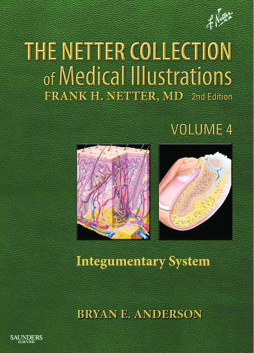
The Netter Collection of Medical Illustrations - Integumentary System E-Book
(2012)
Additional Information
Book Details
Abstract
The Integumentary System, by Bryan E. Anderson, MD, takes a concise and highly visual approach to illustrate the basic sciences and clinical pathology of the skin, hair and nails. This newly added, never-before-published volume in The Netter Collection of Medical Illustrations (formerly the CIBA "Green Books") captures current clinical perspectives on the integumentary system - from normal anatomy and histology to pathology, dermatology, and common issues in plastic surgery and wound healing. Using classic Netter illustrations and new illustrations created in the Netter tradition, as well as a great many cutting-edge histologic micrographs and diagnostic images, it provides a vivid, illuminating, and clinically indispensable view of this body system.
- Gain a rich, holistic clinical view of every structure by seeing classic Netter anatomic illustrations, cutting-edge histologic images and diagnostic imaging studies side by side.
- Visualize the most recent topics in cutaneous pathology such as sporothrix and cutaneous t-cell lymphoma as well as classic problems like alopecia and neurofibromatosis, informed by the latest developments in molecular biology and histologic imaging.
- See current dermatologic concepts captured in the visually rich Netter artistic tradition via major new contributions from Netter disciple Carlos Machado, MD - making complex concepts easy to understand and remember through the precision, clarity, detail, and realism for which Netter’s work has always been known.
- Get complete, integrated visual guidance on the skin, hair, and nails in a single source, from basic sciences and normal anatomy and function through pathologic conditions.
- Adeptly navigate current controversies and timely topics in clinical medicine with guidance from the Editor and informed by an experienced international advisory board.
Table of Contents
| Section Title | Page | Action | Price |
|---|---|---|---|
| Front Cover | cover | ||
| Endsheet | fm1 | ||
| Marketing page p.i | i | ||
| Marketing page p.ii | ii | ||
| The Netter Collection of Medical Illustrations | iii | ||
| Copyright Page | iv | ||
| About the Series | v | ||
| About the Author | vi | ||
| Preface | vii | ||
| About the Artist | viii | ||
| Advisory Board | x | ||
| Table Of Contents | xi | ||
| 1 Anatomy, Physiology, and Embryology | 1 | ||
| Embryology of the Skin | 2 | ||
| Normal Skin Anatomy | 3 | ||
| Normal Skin Histology | 4 | ||
| Skin Physiology: The Process of Keratinization | 5 | ||
| Normal Skin Flora | 6 | ||
| Vitamin D Metabolism | 7 | ||
| Photobiology | 8 | ||
| Wound Healing | 9 | ||
| Morphology | 10 | ||
| 2 Benign Growths | 13 | ||
| Acrochordon | 14 | ||
| Becker’s Nevus (Smooth Muscle Hamartoma) | 15 | ||
| Dermatofibroma (Sclerosing Hemangioma) | 16 | ||
| Eccrine Poroma | 17 | ||
| Eccrine Spiradenoma | 18 | ||
| Eccrine Syringoma | 19 | ||
| Ephelides and Lentigines | 20 | ||
| Epidermal Inclusion Cyst | 22 | ||
| Epidermal Nevus | 23 | ||
| Fibrofolliculoma | 24 | ||
| Fibrous Papule | 25 | ||
| Ganglion Cyst | 26 | ||
| Glomus Tumor and Glomangioma | 27 | ||
| Hidradenoma Papilliferum | 28 | ||
| Hidrocystoma | 29 | ||
| Keloid and Hypertrophic Scar | 30 | ||
| Leiomyoma | 31 | ||
| Lichenoid Keratosis | 32 | ||
| Lipoma | 33 | ||
| Median Raphe Cyst | 34 | ||
| Melanocytic Nevi | 35 | ||
| Milia | 38 | ||
| Neurofibroma | 39 | ||
| Nevus Lipomatosus Superficialis | 40 | ||
| Nevus of Ota and Nevus of Ito | 41 | ||
| Nevus Sebaceus | 42 | ||
| Osteoma Cutis | 43 | ||
| Palisaded Encapsulated Neuroma | 44 | ||
| Pilar Cyst (Trichilemmal Cyst) | 45 | ||
| Porokeratosis | 46 | ||
| Pyogenic Granuloma | 47 | ||
| Reticulohistiocytoma | 48 | ||
| Seborrheic Keratosis | 49 | ||
| Spitz Nevus | 50 | ||
| 3 Malignant Growths | 51 | ||
| Adnexal Carcinoma | 52 | ||
| Angiosarcoma | 53 | ||
| Basal Cell Carcinoma | 54 | ||
| Bowen’s Disease | 56 | ||
| Bowenoid Papulosis | 57 | ||
| Cutaneous Metastases | 58 | ||
| Dermatofibrosarcoma Protuberans | 59 | ||
| Mammary and Extramammary Paget’s Disease | 60 | ||
| Kaposi’s Sarcoma | 61 | ||
| Keratoacanthoma | 62 | ||
| Melanoma | 63 | ||
| Merkel Cell Carcinoma | 65 | ||
| Mycosis Fungoides | 66 | ||
| Sebaceous Carcinoma | 68 | ||
| Squamous Cell Carcinoma | 69 | ||
| 4 Rashes | 71 | ||
| Acanthosis Nigricans | 72 | ||
| Acne | 73 | ||
| Acne Keloidalis Nuchae | 75 | ||
| Acute Febrile Neutrophilic Dermatosis (Sweet’s Syndrome) | 76 | ||
| Allergic Contact Dermatitis | 77 | ||
| Atopic Dermatitis | 79 | ||
| Autoinflammatory Syndromes | 81 | ||
| Bug Bites | 83 | ||
| Calciphylaxis | 85 | ||
| Cutaneous Lupus | 86 | ||
| Cutis Laxa | 89 | ||
| Dermatomyositis | 90 | ||
| Disseminated Intravascular Coagulation | 92 | ||
| Elastosis Perforans Serpiginosa | 93 | ||
| Eruptive Xanthomas | 94 | ||
| Erythema Ab Igne | 96 | ||
| Erythema Annulare Centrifugum | 97 | ||
| Erythema Multiforme, Stevens-Johnson Syndrome, and Toxic Epidermal Necrolysis | 98 | ||
| Erythema Nodosum | 100 | ||
| Fabry Disease | 101 | ||
| Fixed Drug Eruption | 102 | ||
| Gout | 103 | ||
| Graft-Versus-Host Disease | 105 | ||
| Granuloma Annulare | 106 | ||
| Graves’ Disease and Pretibial Myxedema | 107 | ||
| Hidradenitis Suppurativa (Acne Inversa) | 108 | ||
| Irritant Contact Dermatitis | 109 | ||
| Keratosis Pilaris | 110 | ||
| Langerhans Cell Histiocytosis | 111 | ||
| Leukocytoclastic Vasculitis | 113 | ||
| Lichen Planus | 114 | ||
| Lichen Simplex Chronicus | 115 | ||
| Lower Extremity Vascular Insufficiency | 116 | ||
| Mast Cell Disease | 117 | ||
| Morphea | 119 | ||
| Myxedema | 120 | ||
| Necrobiosis Lipoidica | 121 | ||
| Necrobiotic Xanthogranuloma | 122 | ||
| Neutrophilic Eccrine Hidradenitis | 123 | ||
| Ochronosis | 124 | ||
| Oral Manifestations in Blood Dyscrasias | 126 | ||
| Phytophotodermatitis | 127 | ||
| Pigmented Purpura | 128 | ||
| Pityriasis Rosea | 129 | ||
| Pityriasis Rubra Pilaris | 130 | ||
| Polyarteritis Nodosa | 131 | ||
| Pruritic Urticarial Papules and Plaques of Pregnancy | 132 | ||
| Pseudoxanthoma Elasticum | 133 | ||
| Psoriasis | 134 | ||
| Radiation Dermatitis | 137 | ||
| Reactive Arthritis (Reiter’s Syndrome) | 138 | ||
| Rosacea | 139 | ||
| Sarcoid | 140 | ||
| Scleroderma (Progressive Systemic Sclerosis) | 142 | ||
| Seborrheic Dermatitis | 143 | ||
| Skin Manifestations of Inflammatory Bowel Disease | 144 | ||
| Stasis Dermatitis | 146 | ||
| Urticaria | 147 | ||
| Vitiligo | 148 | ||
| 5 Autoimmune Blistering Diseases | 149 | ||
| Basement Membrane Zone, Hemidesmosome, and Desmosome | 150 | ||
| Basement Membrane Zone | 150 | ||
| Hemidesmosome | 151 | ||
| Desmosome | 151 | ||
| Bullous Pemphigoid | 152 | ||
| Mucous Membrane Pemphigoid | 153 | ||
| Dermatitis Herpetiformis | 154 | ||
| Epidermolysis Bullosa Acquisita | 155 | ||
| Linear Immunoglobulin A Bullous Dermatosis | 156 | ||
| Paraneoplastic Pemphigus | 157 | ||
| Pemphigus Foliaceus | 158 | ||
| Pemphigus Vulgaris | 159 | ||
| 6 Infectious Diseases | 161 | ||
| Actinomycosis | 162 | ||
| Blastomycosis | 163 | ||
| Chancroid | 164 | ||
| Coccidioidomycosis | 165 | ||
| Cryptococcosis | 166 | ||
| Cutaneous Larva Migrans | 167 | ||
| Dermatophytoses | 168 | ||
| Herpes Simplex Virus | 171 | ||
| Histoplasmosis | 174 | ||
| Leprosy (Hansen’s Disease) | 175 | ||
| Lice | 176 | ||
| Lyme Disease | 178 | ||
| Lymphogranuloma Venereum | 179 | ||
| Meningococcemia | 180 | ||
| Molluscum Contagiosum | 182 | ||
| Paracoccidioidomycosis | 183 | ||
| Scabies | 184 | ||
| Sporotrichosis | 185 | ||
| Staphylococcus aureus Skin Infections | 186 | ||
| Syphilis | 188 | ||
| Varicella | 191 | ||
| Herpes Zoster (Shingles) | 192 | ||
| Verrucae (Warts) | 194 | ||
| 7 Hair and Nail Diseases | 197 | ||
| Alopecia Areata | 198 | ||
| Androgenic Alopecia | 199 | ||
| Common Nail Disorders | 200 | ||
| Hair Shaft Abnormalities | 203 | ||
| Normal Structure and Function of the Hair Follicle Apparatus | 204 | ||
| Normal Structure and Function of the Nail Unit | 205 | ||
| Telogen Effluvium and Anagen Effluvium | 206 | ||
| Trichotillomania | 207 | ||
| 8 Nutritional and Metabolic Diseases | 209 | ||
| Beriberi | 210 | ||
| Hemochromatosis | 212 | ||
| Metabolic Diseases: Niemann-Pick Disease, von Gierke Disease, and Galactosemia | 213 | ||
| Pellagra | 214 | ||
| Phenylketonuria | 216 | ||
| Scurvy | 218 | ||
| Vitamin A Deficiency | 220 | ||
| Vitamin K Deficiency and Vitamin K Antagonists | 221 | ||
| Wilson’s Disease | 223 | ||
| 9 Genodermatoses and Syndromes | 225 | ||
| Addison’s Disease | 227 | ||
| Amyloidosis | 228 | ||
| Basal Cell Nevus Syndrome | 229 | ||
| Carney Complex | 230 | ||
| Cushing’s Syndrome and Cushing’s Disease | 231 | ||
| Cushing’s Syndrome: Pathophysiology | 232 | ||
| Down Syndrome | 234 | ||
| Ehlers-Danlos Syndrome | 235 | ||
| Marfan Syndrome | 236 | ||
| Neurofibromatosis | 237 | ||
| Tuberous Sclerosis | 239 | ||
| References | 241 | ||
| Section 1: Anatomy, Physiology, and Embryology | 241 | ||
| Section 2: Benign Growths | 241 | ||
| Section 3: Malignant Growths | 242 | ||
| Section 4: Rashes | 242 | ||
| Section 5: Autoimmune Blistering Diseases | 245 | ||
| Section 6: Infectious Diseases | 245 | ||
| Section 7: Hair and Nail Diseases | 247 | ||
| Section 8: Nutritional and Metabolic Diseases | 247 | ||
| Section 9: Genodermatoses and Syndromes | 247 | ||
| Index | 249 | ||
| A | 249 | ||
| B | 249 | ||
| C | 249 | ||
| D | 250 | ||
| E | 250 | ||
| F | 250 | ||
| G | 250 | ||
| H | 251 | ||
| I | 251 | ||
| J | 251 | ||
| K | 252 | ||
| L | 252 | ||
| M | 252 | ||
| N | 252 | ||
| O | 253 | ||
| P | 253 | ||
| Q | 253 | ||
| R | 253 | ||
| S | 254 | ||
| T | 254 | ||
| U | 254 | ||
| V | 254 | ||
| W | 255 | ||
| X | 255 | ||
| Y | 255 | ||
| Z | 255 |
