Additional Information
Book Details
Abstract
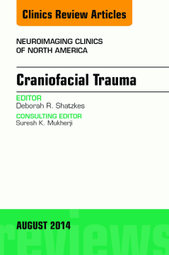
Editor Deborah Shatzkes and authors review important areas in Craniofacial Trauma. Articles will include: Optimizing craniofacial CT technique, Orbital and facial fractures, Orbital and facial soft tissue injuries, Skull base fractures and their complications, Temporal bone trauma, Pediatric considerations in craniofacial trauma, Cerebrovascular trauma, Surgical perspectives in craniofacial trauma, and more!
Table of Contents
| Section Title | Page | Action | Price |
|---|---|---|---|
| Front Cover | Cover | ||
| Craniofacial Trauma\r | i | ||
| Copyright\r | ii | ||
| Neuroimaging Clinics Of North America\r | iv | ||
| Contributors | v | ||
| Contents | vii | ||
| Foreword | xi | ||
| Preface\r | xiii | ||
| Optimizing Craniofacial CT Technique | 395 | ||
| Key points | 395 | ||
| Introduction | 395 | ||
| CT radiation dose measurement | 396 | ||
| CT acquisition parameters | 397 | ||
| Beam Energy | 397 | ||
| Photon Fluence | 397 | ||
| Collimation, Table Speed, and Pitch | 397 | ||
| Current strategies in dose reduction | 398 | ||
| Automated Tube Current Modulation | 398 | ||
| Adaptive Dose Shielding | 398 | ||
| Image Reconstruction Algorithms | 398 | ||
| Future advancement in dose reduction | 399 | ||
| Automated Organ-based Current Modulation | 399 | ||
| Automated (Optimized) Tube Voltage Modulation | 400 | ||
| Noise Reduction Algorithm with Image Reconstruction and Data Processing | 400 | ||
| Three-dimensional Reconstruction | 400 | ||
| CBCT | 400 | ||
| Dual-energy CT | 402 | ||
| Summary | 404 | ||
| References | 404 | ||
| Orbital and Facial Fractures | 407 | ||
| Key points | 407 | ||
| Introduction | 407 | ||
| Frontal sinus | 407 | ||
| Frontal Sinus (Nasofrontal) Outflow Tract | 408 | ||
| Management of Frontal Sinus Fractures | 408 | ||
| Orbital trauma | 409 | ||
| Blow-Out Orbital Fractures | 409 | ||
| Blow-In Orbital Fractures | 410 | ||
| Proptosis and Optic Nerve Injury | 410 | ||
| Complications of Orbital Roof Fractures | 411 | ||
| Cerebrospinal fluid leak | 411 | ||
| Midface fractures | 413 | ||
| Zygomaticomaxillary Complex Fractures | 413 | ||
| Le Fort Fractures | 415 | ||
| Nasal Bones and Septum | 415 | ||
| Naso-Orbito-Ethmoid Fractures | 416 | ||
| Dental | 418 | ||
| Mandible | 419 | ||
| Subcondylar Fractures and Condylar Dislocations | 419 | ||
| Coronoid Process Fractures | 421 | ||
| Summary | 423 | ||
| References | 423 | ||
| Orbital Soft-Tissue Trauma | 425 | ||
| Key points | 425 | ||
| Imaging techniques | 425 | ||
| Orbital anatomy | 425 | ||
| Choice of imaging | 425 | ||
| Radiography | 426 | ||
| Computed Tomography | 426 | ||
| Ultrasonography | 426 | ||
| MR Imaging | 427 | ||
| Anterior chamber injuries | 427 | ||
| Traumatic Hyphema | 427 | ||
| Subconjunctival Hemorrhage | 427 | ||
| Injury to iris and ciliary bodies | 427 | ||
| Lens injuries | 428 | ||
| Subluxation and Dislocation | 428 | ||
| Traumatic Cataract | 428 | ||
| Globe injuries | 429 | ||
| Globe Rupture | 429 | ||
| Globe Luxation | 430 | ||
| Posterior segment injuries | 430 | ||
| Vitreous Hemorrhage | 430 | ||
| Chorioretinal injury | 431 | ||
| Commotio Retinae | 432 | ||
| Penetrating ocular injuries | 432 | ||
| Carotid-cavernous fistulas | 434 | ||
| Optic nerve injury | 434 | ||
| Acknowledgments | 436 | ||
| References | 436 | ||
| Skull Base Fractures and Their Complications | 439 | ||
| Key points | 439 | ||
| Introduction | 439 | ||
| Normal anatomy | 439 | ||
| Pathology | 440 | ||
| Anterior Skull Base Fractures | 442 | ||
| Classification/Etiology | 442 | ||
| Complications | 443 | ||
| Central Skull Base Fractures | 446 | ||
| Classification/Etiology | 446 | ||
| Complications | 447 | ||
| Posterior Skull Base Fractures | 450 | ||
| Classification/Etiology | 450 | ||
| Complications | 451 | ||
| Diagnostic criteria | 453 | ||
| Differential diagnosis | 453 | ||
| Imaging findings | 456 | ||
| Pitfalls in detection of skull base trauma and complications | 459 | ||
| What the referring physician needs to know | 461 | ||
| Future considerations/summary | 462 | ||
| References | 463 | ||
| Imaging of Temporal Bone Trauma | 467 | ||
| Key points | 467 | ||
| Introduction | 467 | ||
| Temporal bone imaging | 467 | ||
| Fracture classification | 468 | ||
| Traditional Classification: Longitudinal and Transverse | 468 | ||
| Alternative Classification Schemes | 468 | ||
| Complications | 470 | ||
| Hearing Loss | 470 | ||
| CHL | 471 | ||
| Ossicular Injury | 471 | ||
| Imaging of ossicular injury | 473 | ||
| Treatment of ossicular injury | 476 | ||
| SNHL | 476 | ||
| Vertigo | 477 | ||
| PLF | 478 | ||
| CSF Leak | 478 | ||
| Imaging findings of CSF leak | 480 | ||
| Treatment of CSF leak | 481 | ||
| Facial Nerve Injury | 482 | ||
| Treatment of facial nerve injury | 482 | ||
| Vascular Injury | 483 | ||
| Summary | 485 | ||
| References | 485 | ||
| Cerebrovascular Trauma | 487 | ||
| Key points | 487 | ||
| Introduction | 487 | ||
| Historical Perspective and Significance of Traumatic Blunt Cerebrovascular Injury | 487 | ||
| Anatomy and pathology | 490 | ||
| Mechanisms and Patterns of Cerebrovascular Injury | 490 | ||
| Pathophysiology of BCVI | 491 | ||
| Imaging | 491 | ||
| Imaging Findings of BCVI | 491 | ||
| BCVI Classification | 494 | ||
| Role of Imaging in BCVI Screening | 496 | ||
| Role of Imaging in BCVI Diagnosis | 501 | ||
| BCVI Treatment and Follow-Up | 505 | ||
| Summary | 509 | ||
| References | 509 | ||
| Pediatric Considerations in Craniofacial Trauma | 513 | ||
| Key points | 513 | ||
| Introduction | 513 | ||
| Normal growth and development | 513 | ||
| Normal variant lucencies in the skull base | 514 | ||
| Normal anterior skull base ossification | 516 | ||
| Paranasal sinus development | 518 | ||
| Distribution and causes of pediatric facial fractures | 519 | ||
| Toppled furniture | 523 | ||
| Nonaccidental, inflicted trauma, and child abuse | 524 | ||
| ATV accidents | 524 | ||
| Impalement injuries | 526 | ||
| Summary | 528 | ||
| References | 528 | ||
| Surgical Perspectives in Craniofacial Trauma | 531 | ||
| Key points | 531 | ||
| Introduction | 531 | ||
| Anatomy | 532 | ||
| Visceral Anatomy | 532 | ||
| Vascular Anatomy | 532 | ||
| Skeletal Anatomy | 532 | ||
| Imaging findings | 533 | ||
| Pathologic abnormality | 534 | ||
| Cranial Injuries | 534 | ||
| Hemorrhagic injuries | 534 | ||
| Intra-axial injury | 536 | ||
| Extra-axial injuries | 536 | ||
| Surgical management of traumatic brain injury | 537 | ||
| Vascular Injuries | 537 | ||
| Skeletal Injuries | 542 | ||
| Skull fractures | 542 | ||
| Frontal sinus fractures | 542 | ||
| Nasal bone fractures | 543 | ||
| Naso-orbital-ethmoidal fractures | 543 | ||
| Injuries to the orbit and orbital contents | 544 | ||
| Fractures of the zygoma and zygomatic-maxillary complex | 545 | ||
| Midface fractures | 545 | ||
| Mandibular fractures | 547 | ||
| Temporal bone fractures | 549 | ||
| Summary | 551 | ||
| Acknowledgments | 551 | ||
| References | 551 | ||
| Index | 553 |
