BOOK
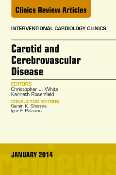
Carotid and Cerebrovascular Disease, An Issue of Interventional Cardiology Clinics, E-Book
Christopher J. White | Kenneth Rosenfield
(2014)
Additional Information
Book Details
Abstract
This issue of Interventional Cardiology Clinics is devoted to Carotid and Cerebrovascular Disease. Expert authors review the most current information available about diagnosing cerebral artery disease and managing carotid and cerebral artery stenosis. Keep up-to-the-minute with the latest developments in cerebrovascular disease interventions.
Table of Contents
| Section Title | Page | Action | Price |
|---|---|---|---|
| Front Cover | Cover | ||
| Carotid and Cerebrovascular Disease\r | i | ||
| Copyright\r | ii | ||
| Contributors | iii | ||
| Contents | v | ||
| Interventional Cardiology Clinics\r | ix | ||
| Preface | xi | ||
| Primary Stroke Prevention | 1 | ||
| Key points | 1 | ||
| Introduction | 1 | ||
| Testing: diagnosis and monitoring | 2 | ||
| Diagnostic Testing | 2 | ||
| Monitoring in Patients with Known Carotid Artery Stenosis | 2 | ||
| Primary prevention interventions | 3 | ||
| Strategies for All Patients | 3 | ||
| Education and lifestyle modification | 3 | ||
| Pharmacologic therapy | 4 | ||
| Antiplatelet therapy | 4 | ||
| Lipid-lowering therapy | 4 | ||
| Blood pressure–lowering therapy | 5 | ||
| Revascularization for primary prevention of stroke | 5 | ||
| Patient Selection and Risk Markers | 5 | ||
| Revascularization Procedures | 6 | ||
| Surgical Carotid Endarterectomy | 6 | ||
| Carotid Stenting | 6 | ||
| Which Procedure to Choose? | 8 | ||
| Summary | 8 | ||
| References | 8 | ||
| Non-Invasive Carotid Imaging | 13 | ||
| Key points | 13 | ||
| Introduction | 13 | ||
| Catheter-based angiography: the historical gold standard | 14 | ||
| Duplex ultrasonography | 14 | ||
| Computed tomography angiography | 15 | ||
| Magnetic resonance angiography | 16 | ||
| Special clinical scenarios | 17 | ||
| Poststent Surveillance | 17 | ||
| Revascularization Planning | 17 | ||
| Recommendations for noninvasive carotid imaging | 18 | ||
| Summary | 19 | ||
| References | 19 | ||
| Skin to Skin | 21 | ||
| Key points | 21 | ||
| Introduction | 21 | ||
| Transradial access technique | 22 | ||
| Transradial carotid angiography | 23 | ||
| Techniques of common carotid artery cannulation | 25 | ||
| Right internal CAS | 26 | ||
| Bovine left internal CAS | 28 | ||
| Nonbovine left internal carotid artery stenting | 28 | ||
| Advanced TRA CAS with Mo.Ma proximal protection device | 31 | ||
| Arterial sheath management | 32 | ||
| Advantages of TRA CAS | 32 | ||
| Disadvantages of TRA CAS | 32 | ||
| Limitations | 32 | ||
| Future technological advances | 33 | ||
| Summary | 33 | ||
| References | 34 | ||
| Skin to Skin | 37 | ||
| Key points | 37 | ||
| Background | 37 | ||
| Preparation of the patient before CAS | 39 | ||
| In-hospital preparation of the CAS patient | 39 | ||
| CAS: access through angiograms | 39 | ||
| CAS: from guide catheter to completion | 40 | ||
| CAS with distal embolic protection | 41 | ||
| Stent selection for CAS | 42 | ||
| Medications during CAS | 43 | ||
| Proximal embolic protection for CAS | 43 | ||
| Theoretical advantages of proximal EPD over distal EPD | 43 | ||
| Relative requirements for proximal EPD | 43 | ||
| Proximal EPD technique during CAS | 44 | ||
| Proximal versus distal EPD decision | 45 | ||
| Common problems encountered during CAS | 45 | ||
| Special situations in CAS | 45 | ||
| Summary | 48 | ||
| References | 48 | ||
| Patient, Anatomic, and Procedural Characteristics That Increase the Risk of Carotid Interventions | 51 | ||
| Key points | 51 | ||
| Introduction | 51 | ||
| Carotid stent risk assessment | 51 | ||
| Patient comorbidities that increase CAS risk | 53 | ||
| Patient Characteristics | 53 | ||
| Symptomatic Versus Asymptomatic Patients | 54 | ||
| Anatomic or lesion characteristics that increase CAS risk | 54 | ||
| Type III Aortic Arch and Aortic Arch Vessel Tortuosity | 54 | ||
| Contralateral Carotid Stenosis or Occlusion | 54 | ||
| Aortic Arch Atheroma | 55 | ||
| Carotid Lesion Characteristics | 55 | ||
| Heavily Calcified Plaque | 55 | ||
| Echolucent Plaque | 55 | ||
| Procedural factors that increase CAS risk | 56 | ||
| Inexperienced Operator and Center | 56 | ||
| Failure to Use an Embolic Protection Device | 56 | ||
| Stent Design | 57 | ||
| Lack of Femoral Artery Access | 57 | ||
| Time to Procedure | 57 | ||
| Summary | 57 | ||
| References | 58 | ||
| Carotid Artery Stenting Versus Carotid Endarterectomy for Treatment of Asymptomatic Carotid Disease | 63 | ||
| Key points | 63 | ||
| Introduction | 63 | ||
| Indications for revascularization of asymptomatic carotid stenosis | 64 | ||
| Randomized trial evidence comparing CEA and CAS in standard-surgical-risk patients | 64 | ||
| Randomized trial evidence comparing CEA and CAS in high-surgical-risk patients | 64 | ||
| Limitations of randomized controlled trial evidence comparing CEA with CAS in asymptomatic patients | 64 | ||
| Observational evidence of CAS in asymptomatic patients | 66 | ||
| Controversies in 2013 | 67 | ||
| CMS Reimbursement | 67 | ||
| Natural History of Asymptomatic Carotid Disease with Optimal Contemporary Medical Therapy | 67 | ||
| Impact of Proximal Embolic Protection on CAS Outcomes | 68 | ||
| Influence of Operator Volume | 68 | ||
| Revascularization of Asymptomatic Disease in the Elderly | 69 | ||
| Importance of Periprocedural Stroke versus MI | 69 | ||
| Deciding therapy for the individual patient | 70 | ||
| Summary | 70 | ||
| References | 70 | ||
| Surgery Versus Stenting in Symptomatic Patients | 73 | ||
| Key points | 73 | ||
| Introduction | 73 | ||
| Patient evaluation | 74 | ||
| Medical treatment | 74 | ||
| Surgical intervention | 75 | ||
| Operative Technique | 77 | ||
| Clinical Trials of Endarterectomy | 78 | ||
| Limitations of Existing Literature | 78 | ||
| Endovascular intervention | 79 | ||
| Interventional Techniques | 79 | ||
| Special Topics in Carotid Stenting | 80 | ||
| Stent design | 80 | ||
| Neuroprotection | 81 | ||
| Clinical Trials of Carotid Stenting | 82 | ||
| Long-term outcomes | 83 | ||
| Summary | 86 | ||
| References | 87 | ||
| Carotid Artery Stenting | 91 | ||
| Key points | 91 | ||
| Introduction | 91 | ||
| CAS learning curve | 92 | ||
| Operator learning curve | 92 | ||
| Institutional Learning Curve | 93 | ||
| Impact of learning curve on randomized control trials | 96 | ||
| How much volume is adequate? | 98 | ||
| Simulation in CAS training | 98 | ||
| Summary | 102 | ||
| References | 102 | ||
| Complications and Solutions with Carotid Stenting | 105 | ||
| Key points | 105 | ||
| Introduction | 105 | ||
| Neurologic complications | 105 | ||
| Cardiovascular complications | 108 | ||
| Death | 109 | ||
| Carotid artery complications | 109 | ||
| Device malfunction | 109 | ||
| General complications | 109 | ||
| Late complications | 109 | ||
| References | 111 | ||
| Percutaneous Treatment of Vertebral Artery Stenosis | 115 | ||
| Key points | 115 | ||
| Introduction | 115 | ||
| Indications and patient selection | 116 | ||
| Relevant anatomy | 116 | ||
| Preprocedure planning | 117 | ||
| Description of procedure | 118 | ||
| Access Site | 118 | ||
| Sheath Sizes | 118 | ||
| Angiographic Catheter | 118 | ||
| Diagnostic Guidewire | 118 | ||
| Interventional Guidewire | 118 | ||
| Percutaneous Transluminal Angioplasty Balloons | 118 | ||
| Stent | 118 | ||
| Interventional Tips | 118 | ||
| Imaging | 118 | ||
| Immediate postprocedural care | 118 | ||
| Clinical results in literature | 119 | ||
| Potential complications/management | 120 | ||
| Summary | 120 | ||
| Videos | 120 | ||
| References | 120 | ||
| Common Cervical and Cerebral Vascular Variants | 123 | ||
| Key points | 123 | ||
| Introduction | 123 | ||
| Anomalies of the aortic arch | 123 | ||
| Normal Anatomy | 123 | ||
| Left-Sided Aortic Arch | 124 | ||
| Common origin of the right brachiocephalic trunk and left common carotid artery | 124 | ||
| Vertebral artery as a direct branch of the aortic arch | 124 | ||
| Aberrant right subclavian artery | 124 | ||
| Other anomalies | 124 | ||
| Right-Sided Aortic Arch | 124 | ||
| Double Aortic Arch | 124 | ||
| Cervical Aortic Arch | 125 | ||
| Anomalies of the common carotid artery | 125 | ||
| Anomalies of the external carotid artery | 125 | ||
| Percutaneous Treatment of Severe Intracranial Carotid and Middle Cerebral Artery Stenosis | 135 | ||
| Key points | 135 | ||
| Introduction | 135 | ||
| Indications and patient selection | 136 | ||
| Cerebrovascular anatomy | 136 | ||
| Preprocedure planning | 137 | ||
| Endovascular approach | 137 | ||
| Perioperative management | 138 | ||
| Clinical results | 138 | ||
| Potential complications and their management | 140 | ||
| Ischemic complications | 140 | ||
| Intracerebral hemorrhage | 141 | ||
| Summary | 141 | ||
| References | 142 | ||
| Current Reperfusion Strategies for Acute Stroke | 145 | ||
| Key points | 145 | ||
| Introduction | 145 | ||
| Indications and patient selection | 146 | ||
| Clinical Status | 146 | ||
| Time of Presentation | 146 | ||
| Imaging of Acute Stroke | 146 | ||
| CT | 147 | ||
| Noncontrast CT | 147 | ||
| CTA | 147 | ||
| CT perfusion | 147 | ||
| MRI | 149 | ||
| Relevant Anatomy | 149 | ||
| Preprocedure planning | 150 | ||
| Optimized Systems-Based Stroke Management | 151 | ||
| Description of the procedure | 152 | ||
| Evolution from IA Thrombolysis to Flow Restoration Devices (Stent Retriever) | 152 | ||
| IA Thrombolysis | 153 | ||
| Merci Device/Penumbra System | 153 | ||
| Flow Restoration Devices/Stent Retriever Technique | 154 | ||
| Stent Retriever Recanalization Technique: Step by Step | 155 | ||
| Vascular access | 155 | ||
| Catheter and sheaths | 155 | ||
| DAC and balloon guide catheter | 156 | ||
| Microwires, microcatheters, and retriever devices | 156 | ||
| IC Recanalization Procedure | 156 | ||
| Technique of the Aspiration Procedure: Step by Step | 158 | ||
| Aspiration indications | 158 | ||
| Aspiration systems | 158 | ||
| Technique | 158 | ||
| Extracranial ICA Occlusion | 158 | ||
| Extracranial revascularization | 160 | ||
| IC Occlusion due to ICA Dissection | 162 | ||
| Potential complications/management | 162 | ||
| Clinical results in the literature | 162 | ||
| Summary | 165 | ||
| Videos | 165 | ||
| References | 165 | ||
| Index | 169 |
