BOOK
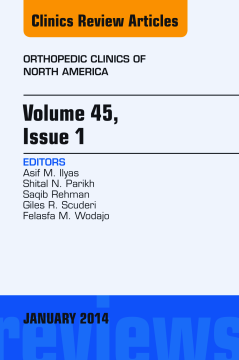
Volume 45, Issue 1, An Issue of Orthopedic Clinics, E-Book
Asif M. Ilyas | Giles R Scuderi | Saqib Rehman | Shital N. Parikh | Felasfa M. Wodajo
(2014)
Additional Information
Book Details
Abstract
Each issue of Orthopedic Clinics offers clinical review articles on the most cutting edge technologies, techniques, and more in the field. Major topic areas include: adult reconstruction, upper extremity, pediatrics, trauma, oncology, hand, foot and ankle, and sports medicine.
Table of Contents
| Section Title | Page | Action | Price |
|---|---|---|---|
| Front Cover | Cover | ||
| Orthopedic Clinics of North America | i | ||
| Copyright | ii | ||
| Contributors | iii | ||
| Contents | vii | ||
| Orthopedic Clinics of North America | xi | ||
| Adult Reconstruction | A1 | ||
| Preface | xiii | ||
| Dual Mobility in Total Hip Arthroplasty | 1 | ||
| Key points | 1 | ||
| Introduction | 1 | ||
| History of dual mobility | 1 | ||
| Biomechanics of dual mobility | 2 | ||
| North American experience | 2 | ||
| Dual mobility in primary THA | 3 | ||
| Dual mobility in revision THA | 5 | ||
| Dual mobility in femoral neck fractures | 7 | ||
| Summary | 7 | ||
| References | 7 | ||
| The Local Effects of Metal Corrosion in Total Hip Arthroplasty | 9 | ||
| Key points | 9 | ||
| Introduction | 9 | ||
| Corrosion at modular interfaces in THA | 9 | ||
| Historical Retrieval Analyses | 10 | ||
| Corrosion in Metal-on-Metal THA | 10 | ||
| Corrosion in Metal-on-Polyethylene THA | 10 | ||
| Corrosion at Neck-Body Junctions | 10 | ||
| Corrosion at Other Modular Junctions | 10 | ||
| Etiology of corrosion | 11 | ||
| Factors Associated with Corrosion | 11 | ||
| Head size | 11 | ||
| Neck length and offset | 11 | ||
| Head material and bearing surface | 11 | ||
| Stem and trunnion design | 12 | ||
| Method of assembly | 12 | ||
| Local effects of metal corrosion in THA | 12 | ||
| Adverse Local Tissue Reactions | 12 | ||
| Instability | 12 | ||
| Loosening and Osteolysis | 13 | ||
| Corrosion-Induced Implant Fracture | 13 | ||
| Remote and Systemic Effects | 13 | ||
| Evaluation and management | 13 | ||
| Diagnostic Workup | 13 | ||
| Treatment Options | 14 | ||
| Summary | 15 | ||
| References | 15 | ||
| The Rationale for Short Uncemented Stems in Total Hip Arthroplasty | 19 | ||
| Key points | 19 | ||
| Introduction | 19 | ||
| Evolution to short stem design | 20 | ||
| Design rationale and types of uncemented short stem metaphyseal-engaging femoral implants | 20 | ||
| Theoretic advantages of short stem femoral implants | 24 | ||
| Ease and Safety of Revision Surgery | 24 | ||
| Revision of a short stem implant | 24 | ||
| Revision of a conventional-length implant | 25 | ||
| Proximal-Distal Mismatch | 26 | ||
| Use in Less-invasive Exposures | 27 | ||
| Clinical and radiographic results of short stem femoral implants | 27 | ||
| Summary | 29 | ||
| References | 30 | ||
| Trauma | A3 | ||
| Preface | xv | ||
| Techniques for Intramedullary Nailing of Proximal Tibia Fractures | 33 | ||
| Key points | 33 | ||
| Introduction | 33 | ||
| Implant design | 34 | ||
| Nail starting point | 34 | ||
| Nailing techniques | 34 | ||
| Nailing in Flexion | 34 | ||
| Nailing in Semiextended Position | 35 | ||
| Suprapatellar Nailing | 36 | ||
| Reduction techniques | 38 | ||
| Clamp-assisted Reduction | 38 | ||
| Blocking/Poller Screws | 38 | ||
| Plate-assisted Reduction | 40 | ||
| Universal Distractor | 44 | ||
| Summary | 44 | ||
| References | 44 | ||
| Perioperative Upper Extremity Peripheral Nerve Traction Injuries | 47 | ||
| Key points | 47 | ||
| Introduction | 47 | ||
| Pathophysiology | 47 | ||
| Classification of peripheral nerve injuries | 48 | ||
| Clinical presentation of peripheral nerve traction injuries | 48 | ||
| General predisposing factors to PPNTI | 48 | ||
| Peripheral nerve traction injuries of the upper limb | 49 | ||
| Status After Shoulder Surgery | 49 | ||
| Brachial plexus injury (C5-T1) | 49 | ||
| Perioperative Lower Extremity Peripheral Nerve Traction Injuries | 55 | ||
| Key points | 55 | ||
| Peripheral nerve traction injuries of the lower limb | 55 | ||
| Following Hip Surgery | 55 | ||
| Sciatic nerve injury (L4–S3) | 56 | ||
| Incidence | 56 | ||
| Predisposing factors | 56 | ||
| Mechanism of injury | 56 | ||
| Clinical presentation | 57 | ||
| Prevention and treatment | 57 | ||
| Pudendal nerve injury (S2–S4) | 58 | ||
| Spondylopelvic Dissociation | 65 | ||
| Key points | 65 | ||
| Spectrum of disease: historical perspective | 65 | ||
| Classification | 66 | ||
| Anatomy and biomechanics | 67 | ||
| Clinical evaluation and related considerations | 67 | ||
| Radiographic assessment | 69 | ||
| Treatment | 70 | ||
| Nonoperative Management | 70 | ||
| Operative Management | 70 | ||
| Soft tissue considerations | 71 | ||
| Fracture alignment and reduction | 71 | ||
| Decompression | 73 | ||
| Instrumentation | 73 | ||
| Surgical outcomes and complications | 74 | ||
| Summary | 74 | ||
| References | 74 | ||
| Pediatrics | A5 | ||
| Preface | xvii | ||
| Slipped Capital Femoral Epiphysis: What’s New? | 77 | ||
| Key points | 77 | ||
| Introduction | 77 | ||
| Prediction of the contralateral SCFE | 78 | ||
| Posterior sloping angle as a predictor of contralateral SCFE | 78 | ||
| The modified Oxford bone age score as a predictor of contralateral SCFE | 79 | ||
| Treatment updates with slipped capital femoral epiphysis | 79 | ||
| Arthrogram-assisted in situ screw fixation | 80 | ||
| Computer navigation–assisted in situ screw fixation | 81 | ||
| Femoral acetabular impingement and acetabular morphology in SCFE | 81 | ||
| Surgical management of femoroacetabular impingement associated with SCFE | 83 | ||
| References | 85 | ||
| Perthes Disease | 87 | ||
| Key points | 87 | ||
| Introduction | 87 | ||
| Natural history | 87 | ||
| Evaluation of prognostic factors | 88 | ||
| Short-term Prognostic Factors | 88 | ||
| Long-term Prognostic Factors | 88 | ||
| Evaluation | 88 | ||
| Patient Evaluation | 88 | ||
| Clinical Evaluation | 88 | ||
| Radiological Evaluation | 88 | ||
| Active stage of disease | 88 | ||
| Early Stage of the Disease | 88 | ||
| Management | 88 | ||
| Containment | 89 | ||
| Nonsurgical containment | 89 | ||
| Surgical containment | 89 | ||
| Type of osteotomy | 89 | ||
| Trochanteric Epiphyseodesis | 90 | ||
| Weight-Bearing Status | 91 | ||
| Late Stage of the Disease | 91 | ||
| Hinge abduction | 91 | ||
| Reducible hinge abduction | 91 | ||
| Irreducible hinge abduction | 91 | ||
| Healed disease | 92 | ||
| Sequelae of perthes disease | 92 | ||
| Morphologic Changes in the Femoral Head | 92 | ||
| Failure of Complete Revacularization | 92 | ||
| Femoroacetabular Impingement | 92 | ||
| Arthritis | 92 | ||
| Clinical Evaluation of Healed Disease | 92 | ||
| Radiological Evaluation of Healed Disease | 92 | ||
| Management of Sequelae | 93 | ||
| Coxa brevis | 93 | ||
| Coxa magna | 93 | ||
| Coxa planna | 93 | ||
| Osteochondritis dessicans | 93 | ||
| FAI | 93 | ||
| Arthritis | 93 | ||
| Evaluation of the outcome at skeletal maturity | 93 | ||
| Future of Perthes | 93 | ||
| References | 94 | ||
| Oncology | A7 | ||
| Preface | xix | ||
| Management of Open Wounds | 99 | ||
| Key points | 99 | ||
| Wound complications in sarcoma treatment | 99 | ||
| Risk factors for poor wound healing | 101 | ||
| Wound vacuum-assisted closure technology | 101 | ||
| Silver topical dressings | 102 | ||
| Mechanism of Silver Action | 102 | ||
| Microbiology of Silver | 103 | ||
| Use of Silver in Open Wounds | 103 | ||
| HBOT | 104 | ||
| Mechanism of Action | 105 | ||
| Hyperbaric Oxygen and Radiation Therapy | 105 | ||
| HBOT and Surgery | 105 | ||
| Other Indications for Use of HBOT | 106 | ||
| Summary | 106 | ||
| References | 106 | ||
| The Practicing Orthopedic Surgeon’s Guide to Managing Long Bone Metastases | 109 | ||
| Key points | 109 | ||
| Introduction | 109 | ||
| Presentation | 109 | ||
| Laboratory workup | 109 | ||
| Imaging workup | 110 | ||
| Pathologic workup | 110 | ||
| Treatments | 111 | ||
| Postoperative management | 114 | ||
| Scenario 1: Painful Bone Lesion in <40 year old | 114 | ||
| Scenario 2: Painful Bone Lesion in ﹥40 Year Old | 114 | ||
| Scenario 3: New Painful Bone Lesion with Remote History of Cancer | 115 | ||
| Scenario 4: Bone Lesion with Current/Recent History of Cancer | 115 | ||
| Scenario 5: Bone Lesion with Impending Pathologic Fracture | 116 | ||
| Scenario 6: Bone Lesion with Pathologic Fracture | 116 | ||
| Scenario 7: Time Distant Metastasis in Renal and Thyroid Cancer | 117 | ||
| Summary | 118 | ||
| References | 118 | ||
| Upper Extremity | A9 | ||
| Preface | xxi | ||
| The Management of Type II Superior Labral Anterior to Posterior Injuries | 121 | ||
| Key points | 121 | ||
| Introduction | 121 | ||
| Causes of failure of SLAP repair | 123 | ||
| The biomechanical and clinical function of the biceps | 124 | ||
| Clinical comparison of biceps tenodesis and SLAP repair | 124 | ||
| Current treatment recommendations | 125 | ||
| References | 126 | ||
| Carpal Instability of the Wrist | 129 | ||
| Key points | 129 | ||
| Introduction | 129 | ||
| Anatomy | 129 | ||
| Biomechanics | 130 | ||
| Mechanism of injury and classification | 130 | ||
| Presentation | 131 | ||
| Examination | 132 | ||
| Studies | 133 | ||
| Treatment | 136 | ||
| Partial scapholunate ligament tears/dynamic instability without diastasis | 136 | ||
| Acute, reducible scapholunate ligament tears | 137 | ||
| Chronic, reducible scapholunate ligament tears | 137 | ||
| Irreducible diastasis/degenerative changes (SLAC wrist) | 139 | ||
| Summary | 139 | ||
| References | 139 | ||
| Kienböck Disease | 141 | ||
| Key points | 141 | ||
| Introduction | 141 | ||
| Cause and pathophysiology | 141 | ||
| Clinical presentation | 142 | ||
| Radiographic imaging | 142 | ||
| Staging | 143 | ||
| Stage I | 143 | ||
| Stage II | 143 | ||
| Stage III | 143 | ||
| Stage IV | 145 | ||
| Treatment | 145 | ||
| Stage I | 146 | ||
| Stage II or IIIA with Negative Ulnar Variance | 146 | ||
| Radial shortening | 146 | ||
| Ulnar shortening | 147 | ||
| Stage II and IIIA with Ulnar Neutral or Positive Variance | 147 | ||
| Revascularization | 147 | ||
| Osteotomies | 147 | ||
| Core decompression | 147 | ||
| Stage IIIB | 148 | ||
| PRC | 148 | ||
| STT or SC arthrodesis | 148 | ||
| Grafting, arthroplasty, interposition | 149 | ||
| Stage IV Disease | 149 | ||
| Summary | 150 | ||
| References | 150 | ||
| Index | 153 |
